31
views
0
recommends
+1 Recommend
0 collections
    0
    shares
      • Record: found
      • Abstract: found
      • Article: found
      Is Open Access

      Risk Factors for Pterygium in Korea: The Korean National Health and Nutrition Examination Survey V, 2010–2012

      research-article
      , PhD, , MD, , MD, PhD, , MD, , MD, PhD
      Medicine
      Wolters Kluwer Health

      Read this article at

      Bookmark
          There is no author summary for this article yet. Authors can add summaries to their articles on ScienceOpen to make them more accessible to a non-specialist audience.

          Abstract

          The aim of this study is to report general and age-specific risk factors for pterygium prevalence in the Korean population.

          This in an observational case series study.

          Data from total 24,812 participants (age 40 years or older) from the Korean National Health and Nutrition Examination Surveys conducted from 2010 to 2012 were retrieved. After applying exclusion criteria, data from 13,204 participants (821 with pterygium and 12,383 without) were used for univariate and multivariate analyses. General risk factors were identified and participants were grouped by decade: 40 s, 50 s, 60 s, 70 s, and 80+. Age-specific risk factors were investigated for each group.

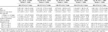
          After univariate analysis, 2 multiple regression models were constructed. Model 1: age + sex + spherical equivalent (SE) + sun exposure hours + occupation (indoor vs outdoor) + residency area (rural vs urban) + education level; model 2: age + sex + SE + sun exposure hours. In model 1, older age (odds ratio [OR]: 1.05 95% confidence interval [CI]: 1.05–1.06), male gender (OR: 1.28, 95% CI: 1.01–1.61), and longer sun exposure hours (OR: 1.47, 95% CI: 1.11–1.94) were significant risk factors for pterygium prevalence whereas higher level of education (elementary school vs college, OR: 3.98, 95% CI: 2.24–7.06) and urban residency (vs rural residency, OR: 0.56, 95% CI: 0.45–0.70) were protective factors. Higher SE (OR 1.11, 95% CI: 1.03–1.19) refractive error was considered a risk factor when using model 2 for the analysis. Age-specific risk factors were different in each age group. Male gender was associated with higher pterygium prevalence in younger age groups while longer sun exposure (5+ hours/day) increased pterygium prevalence in older age groups.

          Previously characterized risk factors were also found in this large population study. However, we found that risk factors may vary according to the age group. Myopic eyes were found to have lower prevalence than hyperopic eyes.

          Related collections

          Most cited references27

          • Record: found
          • Abstract: found
          • Article: not found

          Myopia and level of education: results from the Gutenberg Health Study.

          To analyze the association between myopia and educational level in an adult European cohort. Population-based cross-sectional study. A cohort of the Gutenberg Health Study, including 4658 eligible enrollees between 35 and 74 years of age. We applied a standardized protocol entailing a comprehensive questionnaire; thorough ophthalmic, general, cardiovascular, and psychological examinations; and laboratory tests, including genetic analyses. We documented achievement levels in school education and post-school professional education. The spherical equivalent (SE) was determined by noncycloplegic autorefractometry. We fitted mixed linear models including age, gender, and 45 myopia-associated single nucleotide polymorphisms (SNP) as covariates. Prevalence and magnitude of myopia in association with years spent in school and level of post-school professional education. Individuals who graduated from school after 13 years were more myopic (median, -0.5 diopters [D]; first quartile [Q1]/third quartile [Q3], -2.1/0.3 D) than those who graduated after 10 years (median, -0.2 D; Q1/Q3, -1.3/0.8 D), than those who graduated after 9 years (median, 0.3 D; Q1/Q3, -0.6/1.4 D), and than those who never finished secondary school (median, 0.2 D; Q1/Q3, -0.5/1.8 D; P<0.001, respectively). The same holds true for persons with a university degree (median, -0.6 D; Q1/Q3, -2.3/0.3 D) versus those who finished secondary vocational school (median, 0 D; Q1/Q3, -1.1/0.8 D) or primary vocational school (median, 0 D; Q1/Q3, -0.9/1.1 D) versus persons without any post-school professional qualification (median, 0.6 D; Q1/Q3, -0.4/1.7 D; P<0.001, respectively). Of persons who graduated from school after 13 years, 50.9% were myopic (SE, ≤-0.5 D) versus 41.6%, 27.1%, and 26.9% after 10 years, in those who graduated after 9 years, and in those who never graduated from secondary school, respectively (P<0.001). In university graduates, the proportion of myopic persons was higher (53%) than that of those who graduated from secondary (34.8%) or primary (34.7%) vocational schools and than in those without any professional training (23.9%; P<0.001, respectively). In multivariate analyses: higher school and professional levels of education were associated with a more myopic SE independent of gender. There was a small effect of age and SNPs. Higher levels of school and post-school professional education are associated with a more myopic refraction. Participants with higher educational achievements more often were myopic than individuals with less education. Copyright © 2014 American Academy of Ophthalmology. Published by Elsevier Inc. All rights reserved.
            Bookmark
            • Record: found
            • Abstract: found
            • Article: not found

            Histological changes of high axial myopia.

            J Jonas, L. Xu (2014)
            To describe pathological changes in the anatomy of highly myopic (axially elongated) eyes, enucleated globes were examined by light microscopy and ocular structures were measured histomorphometrically. These studies revealed that highly axially myopic eyes show continuous thinning of the sclera starting at or behind the equator with a maximal thinning at the posterior pole; a profound thinning of the choroid decreasing from ∼ 250 to <10 μm in extreme axial myopia, secondary macular defects in the Bruch's membrane associated with a complete loss of retinal pigment epithelium and choriocapillaris, and retinal photoreceptors; a Bruch's membrane of normal thickness in contrast to the profound thinning of the choroid and the sclera; an up to 10-fold elongation and thinning of the peripapillary scleral flange as anterior roof of the orbital cerebrospinal fluid space, and subsequently a retrobulbar extension of the cerebrospinal fluid space; an increased distance of the peripapillary arterial circle of Zinn-Haller to the optic disc border; an elongation and thinning of the lamina cribrosa with a subsequently decreased distance between the intraocular pressure compartment and the retrobulbar orbital cerebrospinal fluid pressure compartment; an increasing exposure of the peripheral posterior lamina cribrosa surface to the cerebrospinal fluid space, no longer buffered by the solid optic nerve tissue; and the development and enlargement of parapapillary gamma zone, in contrast to a myopia-independent parapapillary beta zone. These anatomical changes may be associated with high axial myopia-related complications such as an increased susceptibility of glaucomatous optic neuropathy and myopic retinopathy.
              Bookmark
              • Record: found
              • Abstract: found
              • Article: not found

              Risk analysis in the development of pterygia.

              No detailed analysis of risk factors in the development of pterygia has been previously performed. In this report, the authors identify and quantify these risk factors. In a case-control study, 278 patients treated in a Brisbane hospital for primary pterygium between 1973 and 1978 were compared with a similar number of people without pterygium who were matched for age, race, and sex, using information from a standard questionnaire completed during a personal interview. Risk of pterygium was increased among patients who, in their third decade of life, worked outdoors in an environment with high surface reflectance of ultraviolet light compared with those who worked indoors. The elevation of risk was raised several hundred-fold among those subjects who worked mainly on sand compared with those who worked indoors when results were adjusted for associated risk factors. When risk was measured among subjects who worked in an environment that was mainly concrete, risk was increased almost 20-fold. Those subjects who spent their first 5 years of life at latitudes less than 30 degrees had almost 40 times the risk of pterygium than those living at latitudes greater than 40 degrees; spending the majority of times outdoors in these earliest years was associated with a 20-fold increase in risk of developing pterygium. There was a strong protective element in the wearing of regular glasses, sunglasses, or a hat. There is a strong suggestion of a causal relationship between ultraviolet light exposure and the development of pterygia during the early years of life and the cumulative exposure over the next 2 to 3 decades in occupations in which there is a high component of reflected ultraviolet light.
                Bookmark

                Author and article information

                Journal
                Medicine (Baltimore)
                Medicine (Baltimore)
                MEDI
                Medicine
                Wolters Kluwer Health
                0025-7974
                1536-5964
                August 2015
                14 August 2015
                : 94
                : 32
                : e1258
                Affiliations
                From the Department of Biostatistics, Dongguk University Ilsan Hospital (C-YL); Department of Ophthalmology, Dongguk University, Ilsan Hospital, Goyang, Kyunggido, South Korea (S-HK, CYP); and Department of Ophthalmology and Visual Sciences, Montefiore Medical Center, Albert Einstein College of Medicine, Bronx, NY, USA (RSC, JKL).
                Author notes
                Correspondence: Choul Y. Park, Department of Ophthalmology, Dongguk University, Ilsan Hospital, 814, Siksadong, Ilsan-dong-gu, Goyang, Kyunggido 410-773, South Korea (e-mail: oph0112@ 123456gmail.com ).
                Article
                01258
                10.1097/MD.0000000000001258
                4616703
                26266358
                af0f53f3-80c4-4d67-8660-8364d099c230
                Copyright © 2015 Wolters Kluwer Health, Inc. All rights reserved.

                This is an open access article distributed under the Creative Commons Attribution-NoDerivatives License 4.0, which allows for redistribution, commercial and non-commercial, as long as it is passed along unchanged and in whole, with credit to the author. http://creativecommons.org/licenses/by-nd/4.0

                History
                : 30 April 2015
                : 4 July 2015
                : 8 July 2015
                Categories
                5800
                Research Article
                Observational Study
                Custom metadata
                TRUE

                Comments

                Comment on this article