27
views
0
recommends
+1 Recommend
0 collections
    0
    shares
      • Record: found
      • Abstract: found
      • Article: found
      Is Open Access

      The Circadian Response of Intrinsically Photosensitive Retinal Ganglion Cells

      research-article

      Read this article at

      Bookmark
          There is no author summary for this article yet. Authors can add summaries to their articles on ScienceOpen to make them more accessible to a non-specialist audience.

          Abstract

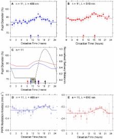
          Intrinsically photosensitive retinal ganglion cells (ipRGC) signal environmental light level to the central circadian clock and contribute to the pupil light reflex. It is unknown if ipRGC activity is subject to extrinsic (central) or intrinsic (retinal) network-mediated circadian modulation during light entrainment and phase shifting. Eleven younger persons (18–30 years) with no ophthalmological, medical or sleep disorders participated. The activity of the inner (ipRGC) and outer retina (cone photoreceptors) was assessed hourly using the pupil light reflex during a 24 h period of constant environmental illumination (10 lux). Exogenous circadian cues of activity, sleep, posture, caffeine, ambient temperature, caloric intake and ambient illumination were controlled. Dim-light melatonin onset (DLMO) was determined from salivary melatonin assay at hourly intervals, and participant melatonin onset values were set to 14 h to adjust clock time to circadian time. Here we demonstrate in humans that the ipRGC controlled post-illumination pupil response has a circadian rhythm independent of external light cues. This circadian variation precedes melatonin onset and the minimum ipRGC driven pupil response occurs post melatonin onset. Outer retinal photoreceptor contributions to the inner retinal ipRGC driven post-illumination pupil response also show circadian variation whereas direct outer retinal cone inputs to the pupil light reflex do not, indicating that intrinsically photosensitive (melanopsin) retinal ganglion cells mediate this circadian variation.

          Related collections

          Most cited references45

          • Record: found
          • Abstract: found
          • Article: not found

          Melanopsin-expressing ganglion cells in primate retina signal colour and irradiance and project to the LGN.

          Human vision starts with the activation of rod photoreceptors in dim light and short (S)-, medium (M)-, and long (L)- wavelength-sensitive cone photoreceptors in daylight. Recently a parallel, non-rod, non-cone photoreceptive pathway, arising from a population of retinal ganglion cells, was discovered in nocturnal rodents. These ganglion cells express the putative photopigment melanopsin and by signalling gross changes in light intensity serve the subconscious, 'non-image-forming' functions of circadian photoentrainment and pupil constriction. Here we show an anatomically distinct population of 'giant', melanopsin-expressing ganglion cells in the primate retina that, in addition to being intrinsically photosensitive, are strongly activated by rods and cones, and display a rare, S-Off, (L + M)-On type of colour-opponent receptive field. The intrinsic, rod and (L + M) cone-derived light responses combine in these giant cells to signal irradiance over the full dynamic range of human vision. In accordance with cone-based colour opponency, the giant cells project to the lateral geniculate nucleus, the thalamic relay to primary visual cortex. Thus, in the diurnal trichromatic primate, 'non-image-forming' and conventional 'image-forming' retinal pathways are merged, and the melanopsin-based signal might contribute to conscious visual perception.
            Bookmark
            • Record: found
            • Abstract: found
            • Article: not found

            Melanopsin and rod-cone photoreceptive systems account for all major accessory visual functions in mice.

            In the mammalian retina, besides the conventional rod-cone system, a melanopsin-associated photoreceptive system exists that conveys photic information for accessory visual functions such as pupillary light reflex and circadian photo-entrainment. On ablation of the melanopsin gene, retinal ganglion cells that normally express melanopsin are no longer intrinsically photosensitive. Furthermore, pupil reflex, light-induced phase delays of the circadian clock and period lengthening of the circadian rhythm in constant light are all partially impaired. Here, we investigated whether additional photoreceptive systems participate in these responses. Using mice lacking rods and cones, we measured the action spectrum for phase-shifting the circadian rhythm of locomotor behaviour. This spectrum matches that for the pupillary light reflex in mice of the same genotype, and that for the intrinsic photosensitivity of the melanopsin-expressing retinal ganglion cells. We have also generated mice lacking melanopsin coupled with disabled rod and cone phototransduction mechanisms. These animals have an intact retina but fail to show any significant pupil reflex, to entrain to light/dark cycles, and to show any masking response to light. Thus, the rod-cone and melanopsin systems together seem to provide all of the photic input for these accessory visual functions.
              Bookmark
              • Record: found
              • Abstract: found
              • Article: not found

              Diminished pupillary light reflex at high irradiances in melanopsin-knockout mice.

              In the mammalian retina, a small subset of retinal ganglion cells (RGCs) are intrinsically photosensitive, express the opsin-like protein melanopsin, and project to brain nuclei involved in non-image-forming visual functions such as pupillary light reflex and circadian photoentrainment. We report that in mice with the melanopsin gene ablated, RGCs retrograde-labeled from the suprachiasmatic nuclei were no longer intrinsically photosensitive, although their number, morphology, and projections were unchanged. These animals showed a pupillary light reflex indistinguishable from that of the wild type at low irradiances, but at high irradiances the reflex was incomplete, a pattern that suggests that the melanopsin-associated system and the classical rod/cone system are complementary in function.
                Bookmark

                Author and article information

                Contributors
                Role: Editor
                Journal
                PLoS One
                plos
                plosone
                PLoS ONE
                Public Library of Science (San Francisco, USA )
                1932-6203
                2011
                14 March 2011
                : 6
                : 3
                : e17860
                Affiliations
                [1 ]Institute of Health and Biomedical Innovation, Queensland University of Technology, Brisbane, Australia
                [2 ]School of Optometry, Queensland University of Technology, Brisbane, Australia
                [3 ]Centre for Accident Research and Road Safety Queensland, Queensland University of Technology, Brisbane, Australia
                University of Houston, United States of America
                Author notes

                Conceived and designed the experiments: AJZ BF SSS. Performed the experiments: ELM AJZ BF SSS. Analyzed the data: ELM AJZ BF SSS. Wrote the paper: AJZ BF SSS.

                Article
                PONE-D-11-01145
                10.1371/journal.pone.0017860
                3056772
                21423755
                b32f4e44-a537-4a12-9e6c-306e7e44f8c2
                Zele et al. This is an open-access article distributed under the terms of the Creative Commons Attribution License, which permits unrestricted use, distribution, and reproduction in any medium, provided the original author and source are credited.
                History
                : 6 January 2011
                : 10 February 2011
                Page count
                Pages: 8
                Categories
                Research Article
                Biology
                Neuroscience
                Sensory Perception
                Psychophysics
                Sensory Systems
                Visual System

                Uncategorized
                Uncategorized

                Comments

                Comment on this article