29
views
0
recommends
+1 Recommend
0 collections
    0
    shares
      • Record: found
      • Abstract: found
      • Article: found
      Is Open Access

      Cinematic rendering – an alternative to volume rendering for 3D computed tomography imaging

      review-article

      Read this article at

      Bookmark
          There is no author summary for this article yet. Authors can add summaries to their articles on ScienceOpen to make them more accessible to a non-specialist audience.

          Abstract

          Abstract

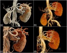
          Volume rendering (VR) represents today’s standard three-dimensional (3-D) image post-processing technique, and often is used to visualize complex anatomical information. Recently, a novel 3-D technique for post-processing of computed tomography (CT) image data has been introduced, which is called cinematic rendering (CR). The objective of this review is to illustrate the image appearance and potential value of CR in comparison with conventional VR in a number of various applications and different anatomical regions. Similar to VR, CR best visualizes high density and high contrast structures such as bones and contrast-enhanced vessels, but at the same time provides a more natural and photo-realistic illumination of the rendered data. Further research will be necessary for determining possible advantages of CR over conventional VR and over two-dimensional (2-D) image post-processing for CT image data.

          Teaching Points

          • Cinematic rendering is a novel post-processing technique for 3D visualization of CT image data .

          • Compared to volume rendering, CR results in a more photo-realistic representation of anatomy .

          • Similar to volume rendering, CR provides best image quality of high density structures.

          Related collections

          Most cited references19

          • Record: found
          • Abstract: found
          • Article: not found

          Three-dimensional volume rendering of spiral CT data: theory and method.

          Three-dimensional (3D) medical images of computed tomographic (CT) data sets can be generated with a variety of computer algorithms. The three most commonly used techniques are shaded surface display, maximum intensity projection, and, more recently, 3D volume rendering. Implementation of 3D volume rendering involves volume data management, which relates to operations including acquisition, resampling, and editing of the data set; rendering parameters including window width and level, opacity, brightness, and percentage classification; and image display, which comprises techniques such as "fly-through" and "fly-around," multiple-view display, obscured structure and shading depth cues, and kinetic and stereo depth cues. An understanding of both the theory and method of 3D volume rendering is essential for accurate evaluation of the resulting images. Three-dimensional volume rendering is useful in a wide variety of applications but is just now being incorporated into commercially available software packages for medical imaging. Although further research is needed to determine the efficacy of 3D volume rendering in clinical applications, with wider availability and improved cost-to-performance ratios in computing, 3D volume rendering is likely to enjoy widespread acceptance in the medical community.
            Bookmark
            • Record: found
            • Abstract: found
            • Article: not found

            Volume rendering versus maximum intensity projection in CT angiography: what works best, when, and why.

            The introduction and widespread availability of 16-section multi-detector row computed tomographic (CT) technology and, more recently, 64-section scanners, has greatly advanced the role of CT angiography in clinical practice. CT angiography has become a key component of state-of-the-art imaging, with applications ranging from oncology (eg, staging of pancreatic or renal cancer) to classic vascular imaging (eg, evaluation of aortic aneurysms and renal artery stenoses) as well as newer techniques such as coronary artery imaging and peripheral runoff studies. With an average of 400-1000 images in each volume data set, three-dimensional postprocessing is crucial to volume visualization. Radiologists now have workstations that provide capabilities for evaluation of these data sets by using a range of software programs and processing tools. Although different systems have unique capabilities and functionality, all provide the options of volume rendering and maximum intensity projection for image display and analysis. These two postprocessing techniques have different advantages and disadvantages when used in clinical practice, and it is important that radiologists understand when and how each technique should be used. Copyright RSNA, 2006.
              Bookmark
              • Record: found
              • Abstract: found
              • Article: found
              Is Open Access

              Prevalence and characteristics of coronary artery anomalies in an adult population undergoing multidetector-row computed tomography for the evaluation of coronary artery disease

              Background Congenital coronary anomalies are uncommon with an incidence ranging from 0.17 % in autopsy cases to 1.2 % in angiographically evaluated cases. The recent development of ECG–gated multi–detector row computed tomography (MDCT) coronary angiography allows accurate and noninvasive depiction of coronary artery anomalies. Methods This retrospective study included 2572 patients who underwent coronary 64-slice MDCT coronary angiography from January 2008 to March 2012. Coronary angiographic scans were obtained with injection of 80 ml nonionic contrast medium. Retrospective gating technique was used to synchronize data reconstruction with the ECG signal. Maximum intensity projection, multi-planar reformatted, and volume rendering images were derived from axial scans. Results Of the 2572 patients, sixty (2.33 %) were diagnosed with coronary artery anomalies (CAAs), with a mean age of 53.6 ± 11.8 years (range 29–80 years). High take-off of the RCA was seen in 16 patients (0.62 %), of the left main coronary artery (LMCA) in 2 patients (0.08 %) and both of them in 2 patients (0.08 %). Separate origin of the left anterior descending artery (LAD) and left circumflex artery (LCx) from left sinus of Valsalva (LSV) was found in 15 patients (an incidence of 0.58 %). In 9 patients (0.35 %) the right coronary artery (RCA) arose from the opposite sinus of Valsalva with a separate ostium. In 6 patients (0.23 %) an abnormal origin of LCX from the right sinus of Valsalva (RSV) was found with a further posterior course within the atrioventricular groove. A single coronary artery was seen in 3 patients (0.12 %). It originated from the right sinus of Valsalva in one patient and from LSV in two patients. In two other patients (0.08 %) the left coronary trunk originated from the RSV with separate ostium from the RCA. LCA originating from the pulmonary artery was found in one patient (0.04 %). A coronary artery fistula, which is a termination anomaly, was detected in 4 patients (0.15 %). Discussion Although these anomalies, which are remarkably different from the normal structure, exist as early as birth, they are incidentally encountered during selective angiography or at autopsy. The incidence in reported angiographic series ranges from 0.6 % to 1.3 %. Variations in the frequency of primary congenital coronary anomalies may possibly have a genetic background. The largest angiographic series of 126595 patients, by Yamanaka and Hobbs, reported a 1.3 % incidence of anomalous coronary artery. Conclusion The results of this study support the use MDCT coronary angiography as a safe and effective noninvasive imaging modality for defining CAAs in an appropriate clinical setting, providing detailed three-dimensional anatomic information that may be difficult to obtain with invasive angiography.
                Bookmark

                Author and article information

                Contributors
                +41 (0) 44 255 3662 , hatem.alkadhi@usz.ch
                Journal
                Insights Imaging
                Insights Imaging
                Insights into Imaging
                Springer Berlin Heidelberg (Berlin/Heidelberg )
                1869-4101
                15 September 2016
                15 September 2016
                December 2016
                : 7
                : 6
                : 849-856
                Affiliations
                [1 ]Institute of Diagnostic and Interventional Radiology, University Hospital Zurich, University of Zurich, Raemistrasse 100, CH- 8091 Zurich, Switzerland
                [2 ]Division of Radiology and Nuclear Medicine, Kantonsspital, St Gallen, Switzerland
                Article
                518
                10.1007/s13244-016-0518-1
                5110476
                27628743
                b5032aa5-f97e-4660-8bcb-a1cfe93ffdfd
                © The Author(s) 2016

                Open Access This article is distributed under the terms of the Creative Commons Attribution 4.0 International License (http://creativecommons.org/licenses/by/4.0/), which permits unrestricted use, distribution, and reproduction in any medium, provided you give appropriate credit to the original author(s) and the source, provide a link to the Creative Commons license, and indicate if changes were made.

                History
                : 6 July 2016
                : 23 August 2016
                : 29 August 2016
                Categories
                Pictorial Review
                Custom metadata
                © The Author(s) 2016

                Radiology & Imaging
                computed tomography,image processing,three-dimensional,volume rendering,cinematic rendering

                Comments

                Comment on this article