1
views
0
recommends
+1 Recommend
0 collections
    0
    shares
      • Record: found
      • Abstract: found
      • Article: found
      Is Open Access

      Mice lacking perforin have improved regeneration of the injured femoral nerve

      research-article

      Read this article at

      Bookmark
          There is no author summary for this article yet. Authors can add summaries to their articles on ScienceOpen to make them more accessible to a non-specialist audience.

          Abstract

          Abstract

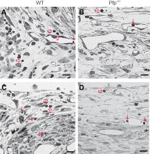
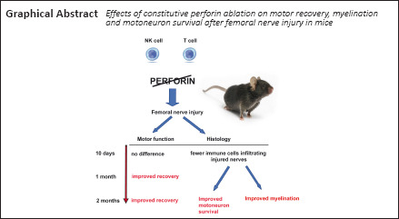
          The role that the immune system plays after injury of the peripheral nervous system is still not completely understood. Perforin, a natural killer cell- and T-lymphocyte-derived enzyme that mediates cytotoxicity, plays important roles in autoimmune diseases, infections and central nervous system trauma, such as spinal cord injury. To dissect the roles of this single component of the immune response to injury, we tested regeneration after femoral nerve injury in perforin-deficient (Pfp –/–) and wild-type control mice. Single frame motion analysis showed better motor recovery in Pfp –/– mice compared with control mice at 4 and 8 weeks after injury. Retrograde tracing of the motoneuron axons regrown into the motor nerve branch demonstrated more correctly projecting motoneurons in the spinal cord of Pfp –/– mice compared with wild-types. Myelination of regrown axons measured by g-ratio was more extensive in Pfp –/– than in wild-type mice in the motor branch of the femoral nerve. Pfp –/– mice displayed more cholinergic synaptic terminals around cell bodies of spinal motoneurons after injury than the injured wild-types. We histologically analyzed lymphocyte infiltration 10 days after surgery and found that in Pfp –/– mice the number of lymphocytes in the regenerating nerves was lower than in wild-types, suggesting a closed blood-nerve barrier in Pfp –/– mice. We conclude that perforin restricts motor recovery after femoral nerve injury owing to decreased survival of motoneurons and reduced myelination.

          Related collections

          Most cited references54

          • Record: found
          • Abstract: found
          • Article: not found

          Perforin and granzymes: function, dysfunction and human pathology.

          A defining property of cytotoxic lymphocytes is their expression and regulated secretion of potent toxins, including the pore-forming protein perforin and serine protease granzymes. Until recently, mechanisms of pore formation and granzyme transfer into the target cell were poorly understood, but advances in structural and cellular biology have now begun to unravel how synergy between perforin and granzymes brings about target cell death. These and other advances are demonstrating the surprisingly broad pathophysiological roles of the perforin–granzyme pathway, and this has important implications for understanding immune homeostasis and for developing immunotherapies for cancer and other diseases. In particular, we are beginning to define and understand a range of human diseases that are associated with a failure to deliver active perforin to target cells. In this Review, we discuss the current understanding of the structural, cellular and clinical aspects of perforin and granzyme biology.
            Bookmark
            • Record: found
            • Abstract: found
            • Article: not found

            The cellular and molecular basis of peripheral nerve regeneration.

            Functional recovery from peripheral nerve injury and repair depends on a multitude of factors, both intrinsic and extrinsic to neurons. Neuronal survival after axotomy is a prerequisite for regeneration and is facilitated by an array of trophic factors from multiple sources, including neurotrophins, neuropoietic cytokines, insulin-like growth factors (IGFs), and glial-cell-line-derived neurotrophic factors (GDNFs). Axotomized neurons must switch from a transmitting mode to a growth mode and express growth-associated proteins, such as GAP-43, tubulin, and actin, as well as an array of novel neuropeptides and cytokines, all of which have the potential to promote axonal regeneration. Axonal sprouts must reach the distal nerve stump at a time when its growth support is optimal. Schwann cells in the distal stump undergo proliferation and phenotypical changes to prepare the local environment to be favorable for axonal regeneration. Schwann cells play an indispensable role in promoting regeneration by increasing their synthesis of surface cell adhesion molecules (CAMs), such as N-CAM, Ng-CAM/L1, N-cadherin, and L2/HNK-1, by elaborating basement membrane that contains many extracellular matrix proteins, such as laminin, fibronectin, and tenascin, and by producing many neurotrophic factors and their receptors. However, the growth support provided by the distal nerve stump and the capacity of the axotomized neurons to regenerate axons may not be sustained indefinitely. Axonal regenerations may be facilitated by new strategies that enhance the growth potential of neurons and optimize the growth support of the distal nerve stump in combination with prompt nerve repair.
              Bookmark
              • Record: found
              • Abstract: found
              • Article: not found

              Macrophage biology in the peripheral nervous system after injury

              Neuroinflammation has positive and negative effects. This review focuses on the roles of macrophage in the PNS. Transection of PNS axons leads to degeneration and clearance of the distal nerve and to changes in the region of the axotomized cell bodies. In both locations, resident and infiltrating macrophages are found. Macrophages enter these areas in response to expression of the chemokine CCL2 acting on the macrophage receptor CCR2. In the distal nerve, macrophages and other phagocytes are involved in clearance of axonal debris, which removes molecules that inhibit nerve regeneration. In the cell body region, macrophage trigger the conditioning lesion response, a process in which neurons increase their regeneration after a prior lesion. In mice in which the genes for CCL2 or CCR2 are deleted, neither macrophage infiltration nor the conditioning lesion response occurs in dorsal root ganglia (DRG). Macrophages exist in different phenotypes depending on their environment. These phenotypes have different effects on axonal clearance and neurite outgrowth. The mechanism by which macrophages affect neuronal cell bodies is still under study. Overexpression of CCL2 in DRG in uninjured animals leads to macrophage accumulation in the ganglia and to an increase in the growth potential of DRG neurons. This increased growth requires activation of neuronal STAT3. In contrast, in acute demyelinating neuropathies, macrophages are involved in stripping myelin from peripheral axons. The molecular mechanisms that trigger macrophage action after trauma and in autoimmune disease are receiving increased attention and should lead to avenues to promote regeneration and protect axonal integrity.
                Bookmark

                Author and article information

                Journal
                Neural Regen Res
                Neural Regen Res
                NRR
                Neural Regeneration Research
                Wolters Kluwer - Medknow (India )
                1673-5374
                1876-7958
                August 2022
                07 January 2022
                : 17
                : 8
                : 1802-1808
                Affiliations
                [1 ]Institut für Anatomie und Klinische Morphologie, Universität Witten/Herdecke, Witten, Germany
                [2 ]Department of Neuroanatomy and Molecular Brain Research, Ruhr University Bochum, Bochum, Germany
                [3 ]Department of Anatomy, Faculty of Medical Sciences, University of Kragujevac, Kragujevac, Serbia
                [4 ]Keck Center for Collaborative Neuroscience and Department of Cell Biology and Neuroscience, Rutgers University, Piscataway, NJ, USA
                Author notes
                [* ] Correspondence to: Melitta Schachner, schachner@ 123456dls.rutgers.edu ; Igor Jakovcevski, Igor.Jakovcevski@ 123456uni-wh.de .

                Author contributions: IJ and MS conceived the study. IJ, MvD, DL, MH and MV performed experiments and collected data. IJ, GR and EF designed experiments and analyzed data. IJ, DL, EF and MS analyzed data and wrote the manuscript. All authors approved the final manuscript version .

                Author information
                https://orcid.org/0000-0002-6654-8402
                https://orcid.org/0000-0002-3316-0778
                Article
                NRR-17-1802
                10.4103/1673-5374.332152
                8820721
                35017441
                b5a94baa-ccd4-4da8-8e40-71052b385535
                Copyright: © Neural Regeneration Research

                This is an open access journal, and articles are distributed under the terms of the Creative Commons Attribution-NonCommercial-ShareAlike 4.0 License, which allows others to remix, tweak, and build upon the work non-commercially, as long as appropriate credit is given and the new creations are licensed under the identical terms.

                History
                : 29 April 2021
                : 07 June 2021
                : 17 July 2021
                Categories
                Research Article

                blood-nerve barrier,femoral nerve injury,locomotor recovery,lymphocytes,myelination,nk-cells,perforin,reinnervation

                Comments

                Comment on this article