5
views
0
recommends
+1 Recommend
0 collections
    0
    shares
      • Record: found
      • Abstract: found
      • Article: found
      Is Open Access

      Area postrema syndrome: Intractable hiccups and vomiting as a result of neuromyelitis Optica Spectrum disorder

      case-report
      1 , , 1
      JRSM Open
      SAGE Publications
      Radiology and imaging, diagnostics, brain stem, cerebellum, neurology

      Read this article at

      Bookmark
          There is no author summary for this article yet. Authors can add summaries to their articles on ScienceOpen to make them more accessible to a non-specialist audience.

          Abstract

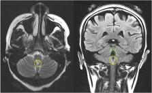
          A 31 year old woman was referred to the medical team for further investigation of intractable hiccups and vomiting. Initial investigations including blood tests, endoscopy and CT imaging did not identify any cause of symptoms. Following multidisciplinary team review, serial MRI Head imaging was arranged, which revealed progressive posterior fossa signal abnormality with involvement of the area postrema. In combination with a positive serum Aquaporin-4 antibody result, this helped establish a diagnosis of Neuromyelitis Optica Spectrum Disorder (NMOSD). Treatment included high dose steroids, plasma exchange and immunomodulatory therapy, and led to a marked improvement in symptoms. This case highlights the importance of utilising specialty team input and broadening lines of investigation, when managing patients with intractable hiccups and vomiting in whom an initial workup has not established a clear diagnosis. While NMOSD is rare, early identification can inform treatment strategies that may lead to a significant improvement in clinical outcome.

          Related collections

          Most cited references12

          • Record: found
          • Abstract: found
          • Article: not found

          International consensus diagnostic criteria for neuromyelitis optica spectrum disorders

          Neuromyelitis optica (NMO) is an inflammatory CNS syndrome distinct from multiple sclerosis (MS) that is associated with serum aquaporin-4 immunoglobulin G antibodies (AQP4-IgG). Prior NMO diagnostic criteria required optic nerve and spinal cord involvement but more restricted or more extensive CNS involvement may occur. The International Panel for NMO Diagnosis (IPND) was convened to develop revised diagnostic criteria using systematic literature reviews and electronic surveys to facilitate consensus. The new nomenclature defines the unifying term NMO spectrum disorders (NMOSD), which is stratified further by serologic testing (NMOSD with or without AQP4-IgG). The core clinical characteristics required for patients with NMOSD with AQP4-IgG include clinical syndromes or MRI findings related to optic nerve, spinal cord, area postrema, other brainstem, diencephalic, or cerebral presentations. More stringent clinical criteria, with additional neuroimaging findings, are required for diagnosis of NMOSD without AQP4-IgG or when serologic testing is unavailable. The IPND also proposed validation strategies and achieved consensus on pediatric NMOSD diagnosis and the concepts of monophasic NMOSD and opticospinal MS.
            Bookmark
            • Record: found
            • Abstract: found
            • Article: not found

            The spectrum of neuromyelitis optica.

            Neuromyelitis optica (also known as Devic's disease) is an idiopathic, severe, demyelinating disease of the central nervous system that preferentially affects the optic nerve and spinal cord. Neuromyelitis optica has a worldwide distribution, poor prognosis, and has long been thought of as a variant of multiple sclerosis; however, clinical, laboratory, immunological, and pathological characteristics that distinguish it from multiple sclerosis are now recognised. The presence of a highly specific serum autoantibody marker (NMO-IgG) further differentiates neuromyelitis optica from multiple sclerosis and has helped to define a neuromyelitis optica spectrum of disorders. NMO-IgG reacts with the water channel aquaporin 4. Data suggest that autoantibodies to aquaporin 4 derived from peripheral B cells cause the activation of complement, inflammatory demyelination, and necrosis that is seen in neuromyelitis optica. The knowledge gained from further assessment of the exact role of NMO-IgG in the pathogenesis of neuromyelitis optica will provide a foundation for rational therapeutic trials for this rapidly disabling disease.
              Bookmark
              • Record: found
              • Abstract: found
              • Article: not found

              Neuromyelitis optica: Evaluation of 871 attacks and 1,153 treatment courses.

              Neuromyelitis optica (NMO) attacks often are severe, are difficult to treat, and leave residual deficits. Here, we analyzed the frequency, sequence, and efficacy of therapies used for NMO attacks.
                Bookmark

                Author and article information

                Journal
                JRSM Open
                JRSM Open
                SHR
                spshr
                JRSM Open
                SAGE Publications (Sage UK: London, England )
                2054-2704
                4 April 2023
                April 2023
                : 14
                : 4
                : 20542704231159601
                Affiliations
                [1 ]Ringgold 4969, universityAcute Medical Unit, Lewisham and Greenwich NHS Trust; , Queen Elizabeth Hospital, Woolwich, London, SE18 4QH, UK
                Author notes
                [*]Kiran Prabhu. Email: kiran.prabhu@ 123456nhs.net
                Author information
                https://orcid.org/0000-0002-8691-0552
                Article
                10.1177_20542704231159601
                10.1177/20542704231159601
                10084536
                b6e5f1b5-1f00-4da0-9349-bed28db68479
                © 2023 The Author(s)

                This article is distributed under the terms of the Creative Commons Attribution-NonCommercial 4.0 License ( https://creativecommons.org/licenses/by-nc/4.0/) which permits non-commercial use, reproduction and distribution of the work without further permission provided the original work is attributed as specified on the SAGE and Open Access page ( https://us.sagepub.com/en-us/nam/open-access-at-sage).

                History
                Categories
                Case Report
                Custom metadata
                ts19

                radiology and imaging,diagnostics,brain stem,cerebellum,neurology

                Comments

                Comment on this article