35
views
0
recommends
+1 Recommend
0 collections
    0
    shares
      • Record: found
      • Abstract: found
      • Article: not found

      c-Jun N-terminal kinase–mediated Rad18 phosphorylation facilitates Polη recruitment to stalled replication forks

      research-article

      Read this article at

      ScienceOpenPublisherPMC
      Bookmark
          There is no author summary for this article yet. Authors can add summaries to their articles on ScienceOpen to make them more accessible to a non-specialist audience.

          Abstract

          The association of Rad18 with Polη is crucial for efficient translesion synthesis and DNA damage tolerance. Rad18–Polη interactions and UV tolerance depend on JNK-dependent Rad18 phosphorylation. These results provide a new mechanism by which SAPK signaling promotes genome maintenance.

          Abstract

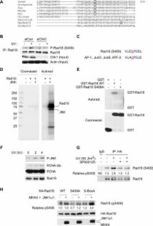
          The E3 ubiquitin ligase Rad18 chaperones DNA polymerase η (Polη) to sites of UV-induced DNA damage and monoubiquitinates proliferating cell nuclear antigen (PCNA), facilitating engagement of Polη with stalled replication forks and promoting translesion synthesis (TLS). It is unclear how Rad18 activities are coordinated with other elements of the DNA damage response. We show here that Ser-409 residing in the Polη-binding motif of Rad18 is phosphorylated in a checkpoint kinase 1–dependent manner in genotoxin-treated cells. Recombinant Rad18 was phosphorylated specifically at S409 by c-Jun N-terminal kinase (JNK) in vitro. In UV-treated cells, Rad18 S409 phosphorylation was inhibited by a pharmacological JNK inhibitor. Conversely, ectopic expression of JNK and its upstream kinase mitogen-activated protein kinase kinase 4 led to DNA damage–independent Rad18 S409 phosphorylation. These results identify Rad18 as a novel JNK substrate. A Rad18 mutant harboring a Ser → Ala substitution at S409 was compromised for Polη association and did not redistribute Polη to nuclear foci or promote Polη−PCNA interaction efficiently relative to wild-type Rad18. Rad18 S409A also failed to fully complement the UV sensitivity of Rad18-depleted cells. Taken together, these results show that Rad18 phosphorylation by JNK represents a novel mechanism for promoting TLS and DNA damage tolerance.

          Related collections

          Most cited references57

          • Record: found
          • Abstract: found
          • Article: not found

          Opposing effects of ERK and JNK-p38 MAP kinases on apoptosis.

          Apoptosis plays an important role during neuronal development, and defects in apoptosis may underlie various neurodegenerative disorders. To characterize molecular mechanisms that regulate neuronal apoptosis, the contributions to cell death of mitogen-activated protein (MAP) kinase family members, including ERK (extracellular signal-regulated kinase), JNK (c-JUN NH2-terminal protein kinase), and p38, were examined after withdrawal of nerve growth factor (NGF) from rat PC-12 pheochromocytoma cells. NGF withdrawal led to sustained activation of the JNK and p38 enzymes and inhibition of ERKs. The effects of dominant-interfering or constitutively activated forms of various components of the JNK-p38 and ERK signaling pathways demonstrated that activation of JNK and p38 and concurrent inhibition of ERK are critical for induction of apoptosis in these cells. Therefore, the dynamic balance between growth factor-activated ERK and stress-activated JNK-p38 pathways may be important in determining whether a cell survives or undergoes apoptosis.
            Bookmark
            • Record: found
            • Abstract: found
            • Article: not found

            Eukaryotic translesion synthesis DNA polymerases: specificity of structure and function.

            This review focuses on eukaryotic translesion synthesis (TLS) DNA polymerases, and the emphasis is on Saccharomyces cerevisiae and human Y-family polymerases (Pols) eta, iota, kappa, and Rev1, as well as on Polzeta, which is a member of the B-family polymerases. The fidelity, mismatch extension ability, and lesion bypass efficiencies of these different polymerases are examined and evaluated in the context of their structures. One major conclusion is that, despite the overall similarity of basic structural features among the Y-family polymerases, there is a high degree of specificity in their lesion bypass properties. Some are able to bypass a particular DNA lesion, whereas others are efficient at only the insertion step or the extension step of lesion bypass. This functional divergence is related to the differences in their structures. Polzeta is a highly specialized polymerase specifically adapted for extending primer termini opposite from a diverse array of DNA lesions, and depending upon the DNA lesion, it contributes to lesion bypass in a mutagenic or in an error-free manner. Proliferating cell nuclear antigen (PCNA) provides the central scaffold to which TLS polymerases bind for access to the replication ensemble stalled at a lesion site, and Rad6-Rad18-dependent protein ubiquitination is important for polymerase exchange.
              Bookmark
              • Record: found
              • Abstract: found
              • Article: not found

              The XPV (xeroderma pigmentosum variant) gene encodes human DNA polymerase eta.

              Xeroderma pigmentosum variant (XP-V) is an inherited disorder which is associated with increased incidence of sunlight-induced skin cancers. Unlike other xeroderma pigmentosum cells (belonging to groups XP-A to XP-G), XP-V cells carry out normal nucleotide-excision repair processes but are defective in their replication of ultraviolet-damaged DNA. It has been suspected for some time that the XPV gene encodes a protein that is involved in trans-lesion DNA synthesis, but the gene product has never been isolated. Using an improved cell-free assay for trans-lesion DNA synthesis, we have recently isolated a DNA polymerase from HeLa cells that continues replication on damaged DNA by bypassing ultraviolet-induced thymine dimers in XP-V cell extracts. Here we show that this polymerase is a human homologue of the yeast Rad30 protein, recently identified as DNA polymerase eta. This polymerase and yeast Rad30 are members of a family of damage-bypass replication proteins which comprises the Escherichia coli proteins UmuC and DinB and the yeast Rev1 protein. We found that all XP-V cells examined carry mutations in their DNA polymerase eta gene. Recombinant human DNA polymerase eta corrects the inability of XP-V cell extracts to carry out DNA replication by bypassing thymine dimers on damaged DNA. Together, these results indicate that DNA polymerase eta could be the XPV gene product.
                Bookmark

                Author and article information

                Contributors
                Role: Monitoring Editor
                Journal
                Mol Biol Cell
                Mol. Biol. Cell
                molbiolcell
                mbc
                Mol. Bio. Cell
                Molecular Biology of the Cell
                The American Society for Cell Biology
                1059-1524
                1939-4586
                15 May 2012
                : 23
                : 10
                : 1943-1954
                Affiliations
                [1] aNational Centre for Biomedical Engineering Science, National University of Ireland, Galway, Ireland
                [2] bDepartment of Pathology and Laboratory Medicine, University of North Carolina, Chapel Hill, NC 27599
                [3] cDana-Farber Cancer Institute, Boston, MA 02215
                [4] dCutaneous Biology Research Center, Massachusetts General Hospital and Harvard Medical School, Charlestown, MA 02129
                [5] eGenome Dynamics Project, Tokyo Metropolitan Institute of Medical Science, 2-1-6 Kamikitazawa, Setagaya-ku, Tokyo 156-8506, Japan
                [6] fCenter for Human Genome Variation, Duke University, Durham, NC 27708
                Vanderbilt University
                Author notes

                *These authors contributed equally to the manuscript.

                1Address correspondence to: Cyrus Vaziri ( cyrus_vaziri@ 123456med.unc.edu ).
                Article
                E11-10-0829
                10.1091/mbc.E11-10-0829
                3350557
                22456510
                b88584e1-b696-458e-8a8c-f4ed1f728f20
                © 2012 Barkley et al. This article is distributed by The American Society for Cell Biology under license from the author(s). Two months after publication it is available to the public under an Attribution–Noncommercial–Share Alike 3.0 Unported Creative Commons License ( http://creativecommons.org/licenses/by-nc-sa/3.0).

                “ASCB®,” “The American Society for Cell Biology®,” and “Molecular Biology of the Cell®” are registered trademarks of The American Society of Cell Biology.

                History
                : 03 October 2011
                : 14 March 2012
                : 19 March 2012
                Categories
                Articles
                Nuclear Functions

                Molecular biology
                Molecular biology

                Comments

                Comment on this article