45
views
0
recommends
+1 Recommend
0 collections
    0
    shares
      • Record: found
      • Abstract: found
      • Article: found
      Is Open Access

      Crystal Structure of Calcium Binding Protein-5 from Entamoeba histolytica and Its Involvement in Initiation of Phagocytosis of Human Erythrocytes

      research-article

      Read this article at

      Bookmark
          There is no author summary for this article yet. Authors can add summaries to their articles on ScienceOpen to make them more accessible to a non-specialist audience.

          Abstract

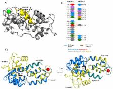
          Entamoeba histolytica is the etiological agent of human amoebic colitis and liver abscess, and causes a high level of morbidity and mortality worldwide, particularly in developing countries. There are a number of studies that have shown a crucial role for Ca 2+ and its binding protein in amoebic biology. EhCaBP5 is one of the EF hand calcium-binding proteins of E. histolytica. We have determined the crystal structure of EhCaBP5 at 1.9 Å resolution in the Ca 2+-bound state, which shows an unconventional mode of Ca 2+ binding involving coordination to a closed yet canonical EF-hand motif. Structurally, EhCaBP5 is more similar to the essential light chain of myosin than to Calmodulin despite its somewhat greater sequence identity with Calmodulin. This structure-based analysis suggests that EhCaBP5 could be a light chain of myosin. Surface plasmon resonance studies confirmed this hypothesis, and in particular showed that EhCaBP5 interacts with the IQ motif of myosin 1B in calcium independent manner. It also appears from modelling of the EhCaBP5-IQ motif complex that EhCaBP5 undergoes a structural change in order to bind the IQ motif of myosin. This specific interaction was further confirmed by the observation that EhCaBP5 and myosin 1B are colocalized in E. histolytica during phagocytic cup formation. Immunoprecipitation of EhCaBP5 from total E. histolytica cellular extract also pulls out myosin 1B and this interaction was confirmed to be Ca 2+ independent. Confocal imaging of E. histolytica showed that EhCaBP5 and myosin 1B are part of phagosomes. Overexpression of EhCaBP5 increases slight rate (∼20%) of phagosome formation, while suppression reduces the rate drastically (∼55%). Taken together, these experiments indicate that EhCaBP5 is likely to be the light chain of myosin 1B. Interestingly, EhCaBP5 is not present in the phagosome after its formation suggesting EhCaBP5 may be playing a regulatory role.

          Author Summary

          Entamoeba histolytica is the etiologic agent of amoebiasis, a major cause of morbidity and mortality in developing countries. The genome of this organism encodes 27 EF-hand containing calcium binding proteins suggesting an intricate Ca 2+ signalling system that plays crucial role in phagocytosis and pathogenesis. Calcium binding protein-5 (EhCaBP5) is one of these CaBPs that displays sequence similarity with Calmodulin (CaM) but has only two possible calcium binding EF-hand loops in two separate domains. Interestingly crystal structure of EhCaPB5 showed more structural similarity with essential light chain (ELC) of myosin than that of CaM. The binding studies of EhCaBP5 with IQ motif peptides of myosins, showed that it interacts with IQ motif of unconventional Myosin IB. A number of experiments were carried out to show that EhCaBP5 indeed binds myosin IB and that this binding is Ca 2+ independent. We also show here that EhCaBP5 participates in erythrophagocytosis and that its role in phagocytosis is different from that of EhCaBP3, another myosin 1B interacting calcium binding protein of E. histolytica. Our results presented here and in a number of other reports point towards a unique phagocytic pathway involving a number of calcium binding proteins in E. histolytica.

          Related collections

          Most cited references31

          • Record: found
          • Abstract: not found
          • Article: not found

          Solvent content of protein crystals.

            Bookmark
            • Record: found
            • Abstract: found
            • Article: not found

            Amoebiasis.

            Amoebiasis is the second leading cause of death from parasitic disease worldwide. The causative protozoan parasite, Entamoeba histolytica, is a potent pathogen. Secreting proteinases that dissolve host tissues, killing host cells on contact, and engulfing red blood cells, E histolytica trophozoites invade the intestinal mucosa, causing amoebic colitis. In some cases amoebas breach the mucosal barrier and travel through the portal circulation to the liver, where they cause abscesses consisting of a few E histolytica trophozoites surrounding dead and dying hepatocytes and liquefied cellular debris. Amoebic liver abscesses grow inexorably and, at one time, were almost always fatal, but now even large abscesses can be cured by one dose of antibiotic. Evidence that what we thought was a single species based on morphology is, in fact, two genetically distinct species--now termed Entamoeba histolytica (the pathogen) and Entamoeba dispar (a commensal)--has turned conventional wisdom about the epidemiology and diagnosis of amoebiasis upside down. New models of disease have linked E histolytica induction of intestinal inflammation and hepatocyte programmed cell death to the pathogenesis of amoebic colitis and amoebic liver abscess.
              Bookmark
              • Record: found
              • Abstract: found
              • Article: not found

              Substructure solution with SHELXD.

              Iterative dual-space direct methods based on phase refinement in reciprocal space and peak picking in real space are able to locate relatively large numbers of anomalous scatterers efficiently from MAD or SAD data. Truncation of the data at a particular resolution, typically in the range 3.0-3.5 A, can be critical to success. The efficiency can be improved by roughly an order of magnitude by Patterson-based seeding instead of starting from random phases or sites; Patterson superposition methods also provide useful validation. The program SHELXD implementing this approach is available as part of the SHELX package.
                Bookmark

                Author and article information

                Contributors
                Role: Editor
                Journal
                PLoS Pathog
                PLoS Pathog
                plos
                plospath
                PLoS Pathogens
                Public Library of Science (San Francisco, USA )
                1553-7366
                1553-7374
                December 2014
                11 December 2014
                : 10
                : 12
                : e1004532
                Affiliations
                [1 ]School of Life Sciences, Jawaharlal Nehru University, New Delhi, India
                [2 ]Department of Biochemistry, Jamia Hamdard, New Delhi, India
                [3 ]Plant Mediator Lab, National Institute of Plant & Genome Research, New Delhi, Delhi, India
                [4 ]European Molecular Biology Laboratory, Grenoble Outstation, France
                [5 ]Unit for Virus Host-Cell Interactions, Université Grenoble Alpes - EMBL-CNRS, France
                University of Virginia Health System, United States of America
                Author notes

                The authors have declared that no competing interests exist.

                Conceived and designed the experiments: SK SA AB SG. Performed the experiments: SK SA MM PD AM BAM. Analyzed the data: SK SA MM BAM RZ AB SG. Contributed reagents/materials/analysis tools: AB SG. Wrote the paper: SK SA MM BAM RZ AB SG.

                Article
                PPATHOGENS-D-14-00103
                10.1371/journal.ppat.1004532
                4263763
                25502654
                b8c301a9-0ca8-4572-9195-82503d4c93b8
                Copyright @ 2014

                This is an open-access article distributed under the terms of the Creative Commons Attribution License, which permits unrestricted use, distribution, and reproduction in any medium, provided the original author and source are credited.

                History
                : 13 January 2014
                : 20 October 2014
                Page count
                Pages: 14
                Funding
                Funding is supported by Department of Biotechnology, Government of India. ( http://www.dbtindia.gov.in/index.asp) The funders had no role in study design, data collection and analysis, decision to publish, or preparation of the manuscript.
                Categories
                Research Article
                Biology and Life Sciences
                Biochemistry
                Proteins
                Protein Interactions
                Protein Structure
                Molecular Biology
                Molecular Complexes

                Infectious disease & Microbiology
                Infectious disease & Microbiology

                Comments

                Comment on this article