14
views
0
recommends
+1 Recommend
0 collections
    0
    shares
      • Record: found
      • Abstract: found
      • Article: found
      Is Open Access

      Association of Alpha-Crystallin with Fiber Cell Plasma Membrane of the Eye Lens Accompanied by Light Scattering and Cataract Formation

      review-article

      Read this article at

      Bookmark
          There is no author summary for this article yet. Authors can add summaries to their articles on ScienceOpen to make them more accessible to a non-specialist audience.

          Abstract

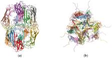
          α-crystallin is a major protein found in the mammalian eye lens that works as a molecular chaperone by preventing the aggregation of proteins and providing tolerance to stress in the eye lens. These functions of α-crystallin are significant for maintaining lens transparency. However, with age and cataract formation, the concentration of α-crystallin in the eye lens cytoplasm decreases with a corresponding increase in the membrane-bound α-crystallin, accompanied by increased light scattering. The purpose of this review is to summarize previous and recent findings of the role of the: (1) lens membrane components, i.e., the major phospholipids (PLs) and sphingolipids, cholesterol (Chol), cholesterol bilayer domains (CBDs), and the integral membrane proteins aquaporin-0 (AQP0; formally MIP26) and connexins, and (2) α-crystallin mutations and post-translational modifications (PTMs) in the association of α-crystallin to the eye lens’s fiber cell plasma membrane, providing thorough insights into a molecular basis of such an association. Furthermore, this review highlights the current knowledge and need for further studies to understand the fundamental molecular processes involved in the association of α-crystallin to the lens membrane, potentially leading to new avenues for preventing cataract formation and progression.

          Related collections

          Most cited references270

          • Record: found
          • Abstract: found
          • Article: not found

          Alpha-crystallin can function as a molecular chaperone.

          J. Horwitz (1992)
          The alpha-crystallins (alpha A and alpha B) are major lens structural proteins of the vertebrate eye that are related to the small heat shock protein family. In addition, crystallins (especially alpha B) are found in many cells and organs outside the lens, and alpha B is overexpressed in several neurological disorders and in cell lines under stress conditions. Here I show that alpha-crystallin can function as a molecular chaperone. Stoichiometric amounts of alpha A and alpha B suppress thermally induced aggregation of various enzymes. In particular, alpha-crystallin is very efficient in suppressing the thermally induced aggregation of beta- and gamma-crystallins, the two other major mammalian structural lens proteins. alpha-Crystallin was also effective in preventing aggregation and in refolding guanidine hydrochloride-denatured gamma-crystallin, as judged by circular dichroism spectroscopy. My results thus indicate that alpha-crystallin refracts light and protects proteins from aggregation in the transparent eye lens and that in nonlens cells alpha-crystallin may have other functions in addition to its capacity to suppress aggregation of proteins.
            Bookmark
            • Record: found
            • Abstract: found
            • Article: found
            Is Open Access

            Mol* Viewer: modern web app for 3D visualization and analysis of large biomolecular structures

            Large biomolecular structures are being determined experimentally on a daily basis using established techniques such as crystallography and electron microscopy. In addition, emerging integrative or hybrid methods (I/HM) are producing structural models of huge macromolecular machines and assemblies, sometimes containing 100s of millions of non-hydrogen atoms. The performance requirements for visualization and analysis tools delivering these data are increasing rapidly. Significant progress in developing online, web-native three-dimensional (3D) visualization tools was previously accomplished with the introduction of the LiteMol suite and NGL Viewers. Thereafter, Mol* development was jointly initiated by PDBe and RCSB PDB to combine and build on the strengths of LiteMol (developed by PDBe) and NGL (developed by RCSB PDB). The web-native Mol* Viewer enables 3D visualization and streaming of macromolecular coordinate and experimental data, together with capabilities for displaying structure quality, functional, or biological context annotations. High-performance graphics and data management allows users to simultaneously visualise up to hundreds of (superimposed) protein structures, stream molecular dynamics simulation trajectories, render cell-level models, or display huge I/HM structures. It is the primary 3D structure viewer used by PDBe and RCSB PDB. It can be easily integrated into third-party services. Mol* Viewer is open source and freely available at https://molstar.org/ . Graphical Abstract Overview of the large array of entities and systems that can be visualized and be manipulated with by the Mol* Viewer.
              Bookmark
              • Record: found
              • Abstract: found
              • Article: not found

              Age-related nuclear cataract-oxidation is the key.

              Age is by far the biggest risk factor for cataract, and it is sometimes assumed that cataract is simply an amplification of this aging process. This appears not to be the case, since the lens changes associated with aging and cataract are distinct. Oxidation is the hallmark of age-related nuclear (ARN) cataract. Loss of protein sulfhydryl groups, and the oxidation of methionine residues, are progressive and increase as the cataract worsens until >90% of cysteine and half the methionine residues are oxidised in the most advanced form. By contrast, there may be no significant oxidation of proteins in the centre of the lens with advancing age, even past age 80. The key factor in preventing oxidation seems to be the concentration of nuclear glutathione (GSH). Provided that nuclear GSH levels can be maintained above 2 mm, it appears that significant protein oxidation and posttranslational modification by reactive small molecules, such as ascorbate or UV filter degradation products, is not observed. Adequate coupling of the metabolically-active cortex, the source of antioxidants such as GSH, to the quiescent nucleus, is crucial especially since it would appear that the cortex remains viable in old lenses, and even possibly in ARN cataract lenses. Therefore it is vital to understand the reason for the onset of the lens barrier. This barrier, which becomes apparent in middle age, acts to impede the flow of small molecules between the cortex and the nucleus. The barrier, rather than nuclear compaction (which is not observed in human lenses), may contribute to the lowered concentration of GSH in the lens nucleus after middle age. By extending the residence time within the lens centre, the barrier also facilitates the decomposition of intrinsically unstable metabolites and may exacerbate the formation of H(2)O(2) in the nucleus. This hypothesis, which is based on the generation of reactive oxygen species and reactive molecules within the nucleus itself, shifts the focus away from theories for cataract that postulated a primary role for oxidants generated outside of the lens. Unfortunately, due to marked variability in the lenses of different species, there appears at present to be no ideal animal model system for studying human ARN cataract.
                Bookmark

                Author and article information

                Contributors
                Role: Academic Editor
                Journal
                Membranes (Basel)
                Membranes (Basel)
                membranes
                Membranes
                MDPI
                2077-0375
                15 June 2021
                June 2021
                : 11
                : 6
                : 447
                Affiliations
                [1 ]Department of Physics, Boise State University, Boise, ID 83725, USA; rajutimsina@ 123456boisestate.edu
                [2 ]Biomolecular Sciences Graduate Program, Boise State University, Boise, ID 83725, USA
                Author notes
                [* ]Correspondence: laxmanmainali@ 123456boisestate.edu ; Tel.: +1-208-426-4003
                Article
                membranes-11-00447
                10.3390/membranes11060447
                8232717
                b98d313c-d959-42b9-ba13-601deb6757f0
                © 2021 by the authors.

                Licensee MDPI, Basel, Switzerland. This article is an open access article distributed under the terms and conditions of the Creative Commons Attribution (CC BY) license ( https://creativecommons.org/licenses/by/4.0/).

                History
                : 24 April 2021
                : 12 June 2021
                Categories
                Review

                α-crystallin,fiber cell plasma membrane,lipids,cholesterol,cholesterol bilayer domains,integral membrane protein,lipid peroxidation,mutations,post-translational modifications,cataract

                Comments

                Comment on this article