4
views
0
recommends
+1 Recommend
0 collections
    0
    shares
      • Record: found
      • Abstract: found
      • Article: found
      Is Open Access

      The use of spectrograms improves the classification of wheezes and crackles in an educational setting

      research-article

      Read this article at

      Bookmark
          There is no author summary for this article yet. Authors can add summaries to their articles on ScienceOpen to make them more accessible to a non-specialist audience.

          Abstract

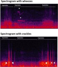
          Chest auscultation is a widely used method in the diagnosis of lung diseases. However, the interpretation of lung sounds is a subjective task and disagreements arise. New technological developments like the use of visSual representation of sounds through spectrograms could improve the agreement when classifying lung sounds, but this is not yet known. In this study, we tested if the use of spectrograms improves the agreement when classifying wheezes and crackles. To do this, we asked twenty-three medical students at UiT the Arctic University of Norway to classify 30 lung sounds recordings for the presence of wheezes and crackles. The sample contained 15 normal recordings and 15 with wheezes or crackles. The students classified the recordings in a random order twice. First sound only, then sound with spectrograms. We calculated kappa values for the agreement between each student and the expert classification with and without display of spectrograms and tested for significant improvement between these two coefficients. We also calculated Fleiss kappa for the 23 observers with and without the spectrogram. In an individual analysis comparing each student to an expert annotated reference standard we found that 13 out of 23 students had a positive change in kappa when classifying wheezes with the help of spectrograms. When classifying crackles 16 out of 23 showed improvement when spectrograms were used. In a group analysis we observed that Fleiss kappa values were k = 0.51 and k = 0.56 (p = 0.63) for classifying wheezes without and with spectrograms. For crackles, these values were k = 0.22 and k = 0.40 (p = <0.01) in the same order. Thus, we conclude that the use of spectrograms had a positive impact on the inter-rater agreement and the agreement with experts. We observed a higher improvement in the classification of crackles compared to wheezes.

          Related collections

          Most cited references19

          • Record: found
          • Abstract: found
          • Article: not found

          Does this patient have community-acquired pneumonia? Diagnosing pneumonia by history and physical examination.

          Community-acquired pneumonia is an important cause of acute respiratory symptoms (eg, cough) in the ambulatory care setting. Distinguishing pneumonia from other causes of respiratory illnesses, such as acute bronchitis and upper respiratory tract infections, has important therapeutic and prognostic implications. The reference standard for diagnosing pneumonia is chest radiography, but it is likely that many physicians rely on the patient's history and their physical examination to diagnose or exclude this disease. A review of published studies of patients suspected of having pneumonia reveals that there are no individual clinical findings, or combinations of findings, that can rule in the diagnosis of pneumonia for a patient suspected of having this illness. However, some studies have shown that the absence of any vital sign abnormalities or any abnormalities on chest auscultation substantially reduces the likelihood of pneumonia to a point where further diagnostic evaluation may be unnecessary. This article reviews the literature on the appropriate use of the history and physical examination in diagnosing community-acquired pneumonia.
            Bookmark
            • Record: found
            • Abstract: found
            • Article: not found

            The rational clinical examination. Does this infant have pneumonia?

            Acute lower respiratory tract illness is common among children seen in primary care. We reviewed the accuracy and precision of the clinical examination in detecting pneumonia in children. Although most cases are viral, it is important to identify bacterial pneumonia to provide appropriate therapy. Studies were identified by searching MEDLINE from 1982 to 1995, reviewing reference lists, reviewing a published compendium of studies of the clinical examination, and consulting experts. Observer agreement is good for most signs on the clinical examination. Each study was reviewed by 2 observers and graded for methodologic quality. There is better agreement about signs that can be observed (eg, use of accessory muscles, color, attentiveness; kappa, 0.48-0.66) than signs that require auscultation of the chest (eg, adventitious sounds; kappa, 0.3). Measurements of the respiratory rate are enhanced by counting for 60 seconds. The best individual finding for ruling out pneumonia is the absence of tachypnea. Chest indrawing, and other signs of increased work of breathing, increases the likelihood of pneumonia. If all clinical signs (respiratory rate, auscultation, and work of breathing) are negative, the chest x-ray findings are unlikely to be positive. Studies are needed to assess the value of clinical findings when they are used together.
              Bookmark
              • Record: found
              • Abstract: not found
              • Article: not found

              Does This Patient Have Community-Acquired Pneumonia?

                Bookmark

                Author and article information

                Contributors
                juan.c.solis@uit.no
                Journal
                Sci Rep
                Sci Rep
                Scientific Reports
                Nature Publishing Group UK (London )
                2045-2322
                21 May 2020
                21 May 2020
                2020
                : 10
                : 8461
                Affiliations
                [1 ]ISNI 0000000122595234, GRID grid.10919.30, General Practice Research Unit, Department of Community Medicine, , UiT The Arctic University of Norway, ; Tromsø, Norway
                [2 ]ISNI 0000000122595234, GRID grid.10919.30, Faculty of Health Sciences, , UiT, The Arctic University of Norway, ; Tromsø, Norway
                [3 ]ISNI 0000 0001 0481 6099, GRID grid.5012.6, Department of methodology and statistics, , University of Maastricht, ; Maastricht, The Netherlands
                Article
                65354
                10.1038/s41598-020-65354-w
                7242373
                32440001
                ba4a4ca2-4fc6-48e9-9c93-e7113b313c37
                © The Author(s) 2020

                Open Access This article is licensed under a Creative Commons Attribution 4.0 International License, which permits use, sharing, adaptation, distribution and reproduction in any medium or format, as long as you give appropriate credit to the original author(s) and the source, provide a link to the Creative Commons license, and indicate if changes were made. The images or other third party material in this article are included in the article’s Creative Commons license, unless indicated otherwise in a credit line to the material. If material is not included in the article’s Creative Commons license and your intended use is not permitted by statutory regulation or exceeds the permitted use, you will need to obtain permission directly from the copyright holder. To view a copy of this license, visit http://creativecommons.org/licenses/by/4.0/.

                History
                : 16 October 2019
                : 24 April 2020
                Categories
                Article
                Custom metadata
                © The Author(s) 2020

                Uncategorized
                physical examination,respiratory signs and symptoms
                Uncategorized
                physical examination, respiratory signs and symptoms

                Comments

                Comment on this article