19
views
0
recommends
+1 Recommend
0 collections
    0
    shares
      • Record: found
      • Abstract: found
      • Article: found
      Is Open Access

      Standard corneal collagen crosslinking versus transepithelial iontophoresis‐assisted corneal crosslinking, 24 months follow‐up: randomized control trial

      research-article
      1 , 2 , , 1
      Acta Ophthalmologica
      John Wiley and Sons Inc.
      crosslinking, iontophoresis, keratoconus, riboflavin

      Read this article at

      Bookmark
          There is no author summary for this article yet. Authors can add summaries to their articles on ScienceOpen to make them more accessible to a non-specialist audience.

          Abstract

          Purpose

          To compare the results of standard corneal crosslinking ( CXL) and transepithelial iontophoresis‐assisted CXL after 24 months follow‐up.

          Material and methods

          Corneal crosslinking (CXL) was performed in a series of 149 eyes of 119 patients with keratoconus I–II of Amsler classification. Depending on the CXL method, patients were divided into two groups: (1) 73 eyes with standard CXL and (2) 76 eyes with transepithelial iontophoresis‐assisted CXL. Depending on the group, epithelium removal or administration of riboflavin solution by iontophoresis for 10 min was performed, after which standard surface UVA irradiation (370 nm, 3 mW/cm 2) was performed at a 5‐cm distance for 30 min.

          Results

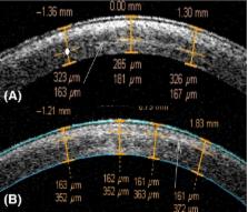
          A statistically significant difference in corrected distance visual acuity ( CDVA) was observed between the two groups, with a better outcome in the second group after 6 months (p = 0.037); however, no significant difference was found 24 months after treatment (p = 0.829). Stabilization and regression of keratometry values were achieved in both groups, but standard CXL was more effective. The average demarcation line depth in the standard CXL group was 292 ± 14  μm after 14 days and 172 ± 16  μm in the transepithelial iontophoresis‐assisted CXL group. No demarcation line was detected after 1 month and 3 months in 45% and 100% of the eyes in the second group respectively.

          Conclusion

          Transepithelial iontophoresis‐assisted collagen crosslinking showed to be less effective than standard CXL after 24 months of follow‐up, possibly due to a more superficial formation of corneal collagen crosslinks, however the stopping of disease progression was achieved 24 months after procedure.

          Related collections

          Most cited references26

          • Record: found
          • Abstract: found
          • Article: not found

          Safety of UVA-riboflavin cross-linking of the cornea.

          To study potential damage to ocular tissue during corneal collagen cross-linking (X-linking) by means of the riboflavin/UVA (370 nm) approach. Comparison of the currently used technique with officially accepted guidelines regarding direct UV damage and the damage created by the induced free radicals (photochemical damage). The currently used UVA radiant exposure of 5.4 mJ/cm and the corresponding irradiance of 3 mW/cm2 is below the known damage thresholds of UVA for the corneal endothelium, lens, and retina. Regarding the photochemical damage caused by the free radicals, the damage thresholds for keratocytes and endothelial cells are 0.45 and 0.35 mW/cm, respectively. In a 400-microm-thick cornea saturated with riboflavin, the irradiance at the endothelial level was 0.18 mW/cm, which is a factor of 2 smaller than the damage threshold. After corneal X-linking, the stroma is depopulated of keratocytes approximately 300 microm deep. Repopulation of this area takes up to 6 months. As long as the cornea treated has a minimum thickness of 400 microm (as recommended), the corneal endothelium will not experience damage, nor will deeper structures such as lens and retina. The light source should provide a homogenous irradiance, avoiding hot spots.
            Bookmark
            • Record: found
            • Abstract: found
            • Article: not found

            Corneal collagen crosslinking with riboflavin and ultraviolet-A light in progressive keratoconus: ten-year results.

            To analyze the 10-year results of corneal collagen crosslinking (CXL) for keratoconus.
              Bookmark
              • Record: found
              • Abstract: found
              • Article: not found

              Biomechanical evidence of the distribution of cross-links in corneas treated with riboflavin and ultraviolet A light.

              To examine to which depth of the cornea the stiffening effect is biomechanically detectable. Department of Ophthalmology, University of Dresden, Dresden, Germany. Of 40 enucleated porcine eyes, 20 eyes were treated with the photosensitizer riboflavin (0.1%) and ultraviolet A (UVA) light (370 nm, 3 mW/cm2, 30 minutes); the other 20 eyes served as control. From each eye, 2 flaps of 200 microm thickness were cut with a microkeratome, and strips of 5 mm width and 7 mm length were prepared. Stress-strain behavior was measured with a material tester to characterize the stiffening effect. Five pairs of human donor eyes were tested in the same way. In porcine corneas, the stiffening effect was stronger in the anterior-treated flaps than in the posterior-treated flaps and the control flaps (P = .001). A 5% strain was achieved at a stress of 261.7 +/- 133.2 x 10(3) N/m2 in the anterior-treated flaps and 104.1 +/- 52.7 x 10(3) N/m2 in the anterior control flaps. The posterior-treated flaps (105.0 +/- 55.8 x 10(3) N/m2) and the posterior control flaps (103.7 +/- 61.8 x 10(3) N/m2) showed no difference (P = .95). A similar stiffening effect was observed in human eyes, but contrary to findings in porcine corneas, in human corneas the anterior control flaps were stiffer than the posterior control flaps (P = .027). Treatment of the cornea with riboflavin and UVA significantly stiffened the cornea only in the anterior 200 microm. This depth-dependent stiffening effect may be explained by the absorption behavior for UVA in the riboflavin-treated cornea. Sixty-five percent to 70% of UVA irradiation was absorbed within the anterior 200 microm and only 20% in the next 200 microm. Therefore, deeper structures and even the endothelium are not affected.
                Bookmark

                Author and article information

                Contributors
                Gbikbova@gmail.com
                Journal
                Acta Ophthalmol
                Acta Ophthalmol
                10.1111/(ISSN)1755-3768
                AOS
                Acta Ophthalmologica
                John Wiley and Sons Inc. (Hoboken )
                1755-375X
                1755-3768
                04 April 2016
                November 2016
                : 94
                : 7 ( doiID: 10.1111/aos.2016.94.issue-7 )
                : e600-e606
                Affiliations
                [ 1 ]Ufa Eye Research Institute UfaRussia
                [ 2 ] Department of Ophthalmology and Visual ScienceChiba University Graduate School of Medicine ChibaJapan
                Author notes
                [*] [* ] Correspondence:

                Guzel Bikbova, MD, PhD

                Department of Ophthalmology and Visual Science

                Chiba University Graduate School of Medicine

                Inohana 1‐8‐1

                Chuo‐ku, Chiba 260‐8670

                Japan

                Tel: +818046316681

                Fax: +810432262005

                Email: Gbikbova@ 123456gmail.com

                Article
                AOS13032
                10.1111/aos.13032
                5111766
                27040458
                bfb944ff-c09d-403e-8824-53f3bbc926be
                © 2016 The Authors. Acta Ophthalmologica published by John Wiley & Sons Ltd on behalf of Acta Ophthalmologica Scandinavica Foundation.

                This is an open access article under the terms of the Creative Commons Attribution‐NonCommercial‐NoDerivs License, which permits use and distribution in any medium, provided the original work is properly cited, the use is non‐commercial and no modifications or adaptations are made.

                History
                : 01 June 2015
                : 22 January 2016
                Page count
                Figures: 4, Tables: 3, Pages: 7, Words: 5162
                Funding
                Funded by: State Academy of Science of Republic Bashkortostan
                Categories
                Original Article
                Original Articles
                Custom metadata
                2.0
                aos13032
                November 2016
                Converter:WILEY_ML3GV2_TO_NLMPMC version:4.9.7 mode:remove_FC converted:16.11.2016

                Ophthalmology & Optometry
                crosslinking,iontophoresis,keratoconus,riboflavin
                Ophthalmology & Optometry
                crosslinking, iontophoresis, keratoconus, riboflavin

                Comments

                Comment on this article