102
views
0
recommends
+1 Recommend
1 collections
    0
    shares
      • Record: found
      • Abstract: found
      • Article: found
      Is Open Access

      Growth Hormone Is Secreted by Normal Breast Epithelium upon Progesterone Stimulation and Increases Proliferation of Stem/Progenitor Cells

      research-article

      Read this article at

      Bookmark
          There is no author summary for this article yet. Authors can add summaries to their articles on ScienceOpen to make them more accessible to a non-specialist audience.

          Summary

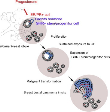
          Using in vitro and in vivo experimental systems and in situ analysis, we show that growth hormone (GH) is secreted locally by normal human mammary epithelial cells upon progesterone stimulation. GH increases proliferation of a subset of cells that express growth hormone receptor (GHR) and have functional properties of stem and early progenitor cells. In 72% of ductal carcinoma in situ lesions, an expansion of the cell population that expresses GHR was observed, suggesting that GH signaling may contribute to breast cancer development.

          Graphical Abstract

          Highlights

          • Growth hormone is produced by normal breast epithelium upon progesterone stimulation

          • Stem/progenitor cells express GHR and proliferate in response to GH stimulation

          • Expansion of GHR-positive cells is seen in precursor lesions of breast malignancies

          Abstract

          Growth hormone (GH) is secreted locally by normal human mammary epithelial cells upon progesterone stimulation. GH signaling increases proliferation of mammary stem and progenitor cells. Analysis of ductal carcinoma in situ lesions shows an expansion of the cell population that expresses GH receptor, suggesting that GH signaling may contribute to breast cancer development.

          Related collections

          Most cited references31

          • Record: found
          • Abstract: found
          • Article: not found

          The tumor suppressor p53 regulates polarity of self-renewing divisions in mammary stem cells.

          Stem-like cells may be integral to the development and maintenance of human cancers. Direct proof is still lacking, mainly because of our poor understanding of the biological differences between normal and cancer stem cells (SCs). Using the ErbB2 transgenic model of breast cancer, we found that self-renewing divisions of cancer SCs are more frequent than their normal counterparts, unlimited and symmetric, thus contributing to increasing numbers of SCs in tumoral tissues. SCs with targeted mutation of the tumor suppressor p53 possess the same self-renewal properties as cancer SCs, and their number increases progressively in the p53 null premalignant mammary gland. Pharmacological reactivation of p53 correlates with restoration of asymmetric divisions in cancer SCs and tumor growth reduction, without significant effects on additional cancer cells. These data demonstrate that p53 regulates polarity of cell division in mammary SCs and suggest that loss of p53 favors symmetric divisions of cancer SCs, contributing to tumor growth.
            Bookmark
            • Record: found
            • Abstract: found
            • Article: not found

            Reconstruction of functionally normal and malignant human breast tissues in mice.

            The study of normal breast epithelial morphogenesis and carcinogenesis in vivo has largely used rodent models. Efforts at studying mammary morphogenesis and cancer with xenotransplanted human epithelial cells have failed to recapitulate the full extent of development seen in the human breast. We have developed an orthotopic xenograft model in which both the stromal and epithelial components of the reconstructed mammary gland are of human origin. Genetic modification of human stromal cells before the implantation of ostensibly normal human mammary epithelial cells resulted in the outgrowth of benign and malignant lesions. This experimental model allows for studies of human epithelial morphogenesis and differentiation in vivo and underscores the critical role of heterotypic interactions in human breast development and carcinogenesis.
              Bookmark
              • Record: found
              • Abstract: found
              • Article: not found

              The role of estrogen in the initiation of breast cancer.

              Estrogens are considered to play a major role in promoting the proliferation of both the normal and the neoplastic breast epithelium. Their role as breast carcinogens has long been suspected and recently confirmed by epidemiological studies. Three major mechanisms are postulated to be involved in their carcinogenic effects: stimulation of cellular proliferation through their receptor-mediated hormonal activity, direct genotoxic effects by increasing mutation rates through a cytochrome P450-mediated metabolic activation, and induction of aneuploidy. Recently it has been fully demonstrated that estrogens are carcinogenic in the human breast by testing in an experimental system the natural estrogen 17beta-estradiol (E(2)) by itself or its metabolites 2-hydroxy, 4-hydroxy, and 16-a-hydroxy-estradiol (2-OH-E(2), 4-OH-E(2), and 16-alpha-OH E(2)), respectively, by inducing neoplastic transformation of human breast epithelial cells (HBEC) MCF-10F in vitro to a degree at least similar to that induced by the chemical carcinogen benz(a)pyrene (BP). Neither Tamoxyfen (TAM) nor ICI-182,780 abrogated the transforming efficiency of estrogen or its metabolites. The E(2) induced expression of anchorage independent growth, loss of ductulogenesis in collagen, invasiveness in Matrigel, is associated with the loss of 9p11-13 and only invasive cells that exhibited a 4p15.3-16 deletion were tumorigenic. Tumors were poorly differentiated ER-alpha and progesterone receptor negative adenocarcinomas that expressed keratins, EMA and E-cadherin. The E(2) induced tumors and tumor-derived cell lines exhibited loss of chromosome 4, deletions in chromosomes 3p12.3-13, 8p11.1-21, 9p21-qter, and 18q, and gains in 1p, and 5q15-qter. The induction of complete transformation of the human breast epithelial cell MCF-10F in vitro confirms the carcinogenicity of E(2), supporting the concept that this hormone could act as an initiator of breast cancer in women. This model provides a unique system for understanding the genomic changes that intervene for leading normal cells to tumorigenesis and for testing the functional role of specific genomic events taking place during neoplastic transformation.
                Bookmark

                Author and article information

                Contributors
                Journal
                Stem Cell Reports
                Stem Cell Reports
                Stem Cell Reports
                Elsevier
                2213-6711
                3 June 2014
                3 June 2014
                03 June 2014
                : 2
                : 6
                : 780-793
                Affiliations
                [1 ]Research Oncology, King’s College London, London SE1 9RT, UK
                [2 ]Centre de Recherche et Cancérologie, Marseille, Inserm, CRCM, U1068, France
                [3 ]Department of Internal Medicine, University of Michigan, Ann Arbor, MI 48109, USA
                Author notes
                []Corresponding author gabriela.dontu@ 123456kcl.ac.uk
                Article
                S2213-6711(14)00146-5
                10.1016/j.stemcr.2014.05.005
                4050343
                24936466
                bfed9e52-4464-4386-bb53-51f07edfa7c8
                © 2014 The Authors

                This is an open access article under the CC BY-NC-ND license (http://creativecommons.org/licenses/by-nc-nd/3.0/).

                History
                : 17 April 2013
                : 6 May 2014
                : 7 May 2014
                Categories
                Article

                Comments

                Comment on this article