10
views
0
recommends
+1 Recommend
0 collections
    0
    shares
      • Record: found
      • Abstract: found
      • Article: found
      Is Open Access

      Vibratory stimulation enhances thyroid epithelial cell function

      research-article

      Read this article at

      Bookmark
          There is no author summary for this article yet. Authors can add summaries to their articles on ScienceOpen to make them more accessible to a non-specialist audience.

          Abstract

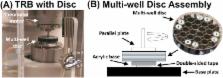
          The tissues of the body are routinely subjected to various forms of mechanical vibration, the frequency, amplitude, and duration of which can contribute both positively and negatively to human health. The vocal cords, which are in close proximity to the thyroid, may also supply the thyroid with important mechanical signals that modulate hormone production via mechanical vibrations from phonation. In order to explore the possibility that vibrational stimulation from vocalization can enhance thyroid epithelial cell function, FRTL-5 rat thyroid cells were subjected to either chemical stimulation with thyroid stimulating hormone (TSH), mechanical stimulation with physiological vibrations, or a combination of the two, all in a well-characterized, torsional rheometer-bioreactor. The FRTL-5 cells responded to mechanical stimulation with significantly (p<0.05) increased metabolic activity, significantly (p<0.05) increased ROS production, and increased gene expression of thyroglobulin and sodium-iodide symporter compared to un-stimulated controls, and showed an equivalent or greater response than TSH only stimulated cells. Furthermore, the combination of TSH and oscillatory motion produced a greater response than mechanical or chemical stimulation alone. Taken together, these results suggest that mechanical vibrations could provide stimulatory cues that help maintain thyroid function.

          Highlights

          • Thyroid epithelial cells responded to mechanical vibrations similar to those from vocalization.

          • This response was equivalent or greater compared to chemical stimulation.

          • The combination of mechanical and chemical stimulation was synergistic.

          • It may be possible to influence thyroid function with mechanical vibrations.

          Related collections

          Most cited references57

          • Record: found
          • Abstract: found
          • Article: not found

          Quantifying cellular oxidative stress by dichlorofluorescein assay using microplate reader.

          Oxidative stress (OS) has been implicated in various degenerative diseases in aging. In an attempt to quantify OS in a cell model, we examined OS induced by incubating for 30 min with various free radical generators in PC12 cells by using the dichlorofluorescein (DCF) assay, modified for use by a fluorescent microplate reader. The nonfluorescent fluorescin derivatives (dichlorofluorescin, DCFH), after being oxidized by various oxidants, will become DCF and emit fluorescence. By quantifying the fluorescence, we were able to quantify the OS. Our results indicated that the fluorescence varied linearly with increasing concentrations (between 0.1 and 1 mM) of H2O2 and 2,2'-azobios(2-amidinopropane) dihydrochloride (AAPH; a peroxyl radical generator). By contrast, the fluorescence varied as a nonlinear response to increasing concentrations of 3-morpholinosydnonimine hydrochloride (SIN-1; a peroxynitrite generator), sodium nitroprusside (SNP; a nitric oxide generator), and dopamine. Dopamine had a biphasic effect; it decreased the DCF fluorescence, thus acting as an antioxidant, at concentrations <500 microM in cells, but acted as a pro-oxidant by increasing the fluorescence at 1 mM. While SNP was not a strong pro-oxidant, SIN-1 was the most potent pro-oxidant among those tested, inducing a 70 times increase of fluorescence at a concentration of 100 microM compared with control. Collectively, due to its indiscriminate nature to various free radicals, DCF can be very useful in quantifying overall OS in cells, especially when used in conjunction with a fluorescent microplate reader. This method is reliable and efficient for evaluating the potency of pro-oxidants and can be used to evaluate the efficacy of antioxidants against OS in cells.
            Bookmark
            • Record: found
            • Abstract: found
            • Article: not found

            Increasing incidence of differentiated thyroid cancer in the United States, 1988-2005.

            Studies have reported an increasing incidence of thyroid cancer since 1980. One possible explanation for this trend is increased detection through more widespread and aggressive use of ultrasound and image-guided biopsy. Increases resulting from increased detection are most likely to involve small primary tumors rather than larger tumors, which often present as palpable thyroid masses. The objective of the current study was to investigate the trends in increasing incidence of differentiated (papillary and follicular) thyroid cancer by size, age, race, and sex. Cases of differentiated thyroid cancer (1988-2005) were analyzed using the National Cancer Institute's Surveillance Epidemiology and End Results (SEER) dataset. Trends in incidence rates of papillary and follicular cancer, race, age, sex, primary tumor size ( 4 cm), and SEER stage (localized, regional, distant) were analyzed using joinpoint regression and reported as the annual percentage change (APC). Incidence rates increased for all sizes of tumors. Among men and women of all ages, the highest rate of increase was for primary tumors or =4 cm among men (1988-2005: APC, 3.7) and women (1988-2005: APC, 5.70) and for distant SEER stage disease among men (APC, 3.7) and women (APC, 2.3). The incidence rates of differentiated thyroid cancers of all sizes increased between 1988 and 2005 in both men and women. The increased incidence across all tumor sizes suggested that increased diagnostic scrutiny is not the sole explanation. Other explanations, including environmental influences and molecular pathways, should be investigated.
              Bookmark
              • Record: found
              • Abstract: found
              • Article: not found

              Regulation of thyroid cell proliferation by TSH and other factors: a critical evaluation of in vitro models.

              TSH via cAMP, and various growth factors, in cooperation with insulin or IGF-I stimulate cell cycle progression and proliferation in various thyrocyte culture systems, including rat thyroid cell lines (FRTL-5, WRT, PC Cl3) and primary cultures of rat, dog, sheep and human thyroid. The available data on cell signaling cascades, cell cycle kinetics, and cell cycle-regulatory proteins are thoroughly and critically reviewed in these experimental systems. In most FRTL-5 cells, TSH (cAMP) merely acts as a priming/competence factor amplifying PI3K and MAPK pathway activation and DNA synthesis elicited by insulin/IGF-I. In WRT cells, TSH and insulin/IGF-I can independently activate Ras and PI3K pathways and DNA synthesis. In dog thyroid primary cultures, TSH (cAMP) does not activate Ras and PI3K, and cAMP must be continuously elevated by TSH to directly control the progression through G(1) phase. This effect is exerted, at least in part, via the cAMP-dependent activation of the required cyclin D3, itself synthesized in response to insulin/IGF-I. This and other discrepancies show that the mechanistic logics of cell cycle stimulation by cAMP profoundly diverge in these different in vitro models of the same cell. Therefore, although these different thyrocyte systems constitute interesting models of the wide diversity of possible mechanisms of cAMP-dependent proliferation in various cell types, extrapolation of in vitro mechanistic data to TSH-dependent goitrogenesis in man can only be accepted in the cases where independent validation is provided.
                Bookmark

                Author and article information

                Contributors
                Journal
                Biochem Biophys Rep
                Biochem Biophys Rep
                Biochemistry and Biophysics Reports
                Elsevier
                2405-5808
                19 October 2016
                December 2016
                19 October 2016
                : 8
                : 376-381
                Affiliations
                [a ]Department of Biomedical Engineering, University of Iowa, IA, USA
                [b ]Department of Communication Sciences and Disorders, University of Iowa, IA, USA
                [c ]National Center for Voice and Speech, University of Utah, Salt Lake City, UT, USA
                Author notes
                [* ]Correspondence to: Department of Biomedical Engineering, University of Iowa, 1402 Seamans Center, Iowa City, IA 52242, USA. edward-sander@ 123456uiowa.edu
                Article
                S2405-5808(16)30222-9
                10.1016/j.bbrep.2016.10.008
                5614476
                28955979
                c0155e74-0eb6-4f91-8aeb-c322626c768d
                © 2016 The Authors

                This is an open access article under the CC BY-NC-ND license (http://creativecommons.org/licenses/by-nc-nd/4.0/).

                History
                : 13 April 2016
                : 11 October 2016
                : 13 October 2016
                Categories
                Research Article

                frtl-5,mechanobiology,mechanotransduction,low amplitude mechanical signals,endocrine system

                Comments

                Comment on this article