5
views
0
recommends
+1 Recommend
0 collections
    0
    shares
      • Record: found
      • Abstract: found
      • Article: found
      Is Open Access

      Identification of hepatic fibrosis inhibitors through morphometry analysis of a hepatic multicellular spheroids model

      research-article

      Read this article at

      Bookmark
          There is no author summary for this article yet. Authors can add summaries to their articles on ScienceOpen to make them more accessible to a non-specialist audience.

          Abstract

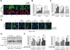
          A chronic, local inflammatory milieu can cause tissue fibrosis that results in epithelial-to-mesenchymal transition (EMT), endothelial-to-mesenchymal transition (EndMT), increased abundance of fibroblasts, and further acceleration of fibrosis. In this study, we aimed to identify potential mechanisms and inhibitors of fibrosis using 3D model-based phenotypic screening. We established liver fibrosis models using multicellular tumor spheroids (MCTSs) composed of hepatocellular carcinoma (HCC) and stromal cells such as fibroblasts (WI38), hepatic stellate cells (LX2), and endothelial cells (HUVEC) seeded at constant ratios. Through high-throughput screening of FDA-approved drugs, we identified retinoic acid and forskolin as candidates to attenuate the compactness of MCTSs as well as inhibit the expression of ECM-related proteins. Additionally, retinoic acid and forskolin induced reprogramming of fibroblast and cancer stem cells in the HCC microenvironment. Of interest, retinoic acid and forskolin had anti-fibrosis effects by decreasing expression of α-SMA and F-actin in LX2 cells and HUVEC cells. Moreover, when sorafenib was added along with retinoic acid and forskolin, apoptosis was increased, suggesting that anti-fibrosis drugs may improve tissue penetration to support the efficacy of anti-cancer drugs. Collectively, these findings support the potential utility of morphometric analyses of hepatic multicellular spheroid models in the development of new drugs with novel mechanisms for the treatment of hepatic fibrosis and HCCs.

          Related collections

          Most cited references46

          • Record: found
          • Abstract: found
          • Article: not found

          Liver fibrosis.

          Liver fibrosis is the excessive accumulation of extracellular matrix proteins including collagen that occurs in most types of chronic liver diseases. Advanced liver fibrosis results in cirrhosis, liver failure, and portal hypertension and often requires liver transplantation. Our knowledge of the cellular and molecular mechanisms of liver fibrosis has greatly advanced. Activated hepatic stellate cells, portal fibroblasts, and myofibroblasts of bone marrow origin have been identified as major collagen-producing cells in the injured liver. These cells are activated by fibrogenic cytokines such as TGF-beta1, angiotensin II, and leptin. Reversibility of advanced liver fibrosis in patients has been recently documented, which has stimulated researchers to develop antifibrotic drugs. Emerging antifibrotic therapies are aimed at inhibiting the accumulation of fibrogenic cells and/or preventing the deposition of extracellular matrix proteins. Although many therapeutic interventions are effective in experimental models of liver fibrosis, their efficacy and safety in humans is unknown. This review summarizes recent progress in the study of the pathogenesis and diagnosis of liver fibrosis and discusses current antifibrotic strategies.
            Bookmark
            • Record: found
            • Abstract: found
            • Article: found
            Is Open Access

            Three-Dimensional in Vitro Cell Culture Models in Drug Discovery and Drug Repositioning

            Drug development is a lengthy and costly process that proceeds through several stages from target identification to lead discovery and optimization, preclinical validation and clinical trials culminating in approval for clinical use. An important step in this process is high-throughput screening (HTS) of small compound libraries for lead identification. Currently, the majority of cell-based HTS is being carried out on cultured cells propagated in two-dimensions (2D) on plastic surfaces optimized for tissue culture. At the same time, compelling evidence suggests that cells cultured in these non-physiological conditions are not representative of cells residing in the complex microenvironment of a tissue. This discrepancy is thought to be a significant contributor to the high failure rate in drug discovery, where only a low percentage of drugs investigated ever make it through the gamut of testing and approval to the market. Thus, three-dimensional (3D) cell culture technologies that more closely resemble in vivo cell environments are now being pursued with intensity as they are expected to accommodate better precision in drug discovery. Here we will review common approaches to 3D culture, discuss the significance of 3D cultures in drug resistance and drug repositioning and address some of the challenges of applying 3D cell cultures to high-throughput drug discovery.
              Bookmark
              • Record: found
              • Abstract: found
              • Article: not found

              Drug penetration in solid tumours.

              To be most effective anticancer drugs must penetrate tissue efficiently, reaching all the cancer cells that comprise the target population in a concentration sufficient to exert a therapeutic effect. Most research into the resistance of cancers to chemotherapy has concentrated on molecular mechanisms of resistance, whereas the role of limited drug distribution within tumours has been neglected. We summarize the evidence that indicates that the distribution of many anticancer drugs in tumour tissue is incomplete, and we suggest strategies that might be used either to improve drug penetration through tumour tissue or to select compounds based on their abilities to penetrate tissue, thereby increasing the therapeutic index.
                Bookmark

                Author and article information

                Contributors
                shr1261@ip-korea.org
                Journal
                Sci Rep
                Sci Rep
                Scientific Reports
                Nature Publishing Group UK (London )
                2045-2322
                25 May 2021
                25 May 2021
                2021
                : 11
                : 10931
                Affiliations
                [1 ]GRID grid.418549.5, ISNI 0000 0004 0494 4850, Cancer Biology Laboratory, , Institut Pasteur Korea, ; 16, Daewangpangyo-ro 712 beon-gil, Bundang-gu, Seongnam-si, 13488 Gyeonggi-do Republic of Korea
                [2 ]GRID grid.418549.5, ISNI 0000 0004 0494 4850, Screening Discovery Platform, , Institut Pasteur Korea, ; 16, Daewangpangyo-ro 712 beon-gil, Bundang-gu, Seongnam-si, 13488 Gyeonggi-do Republic of Korea
                [3 ]GRID grid.412786.e, ISNI 0000 0004 1791 8264, Division of Bio-Medical Science and Technology, , University of Science and Technology, ; Deajeon, 34113 Republic of Korea
                Article
                90263
                10.1038/s41598-021-90263-x
                8149639
                34035369
                c217bb30-18b2-404d-9a03-cec4e34f2207
                © The Author(s) 2021

                Open Access This article is licensed under a Creative Commons Attribution 4.0 International License, which permits use, sharing, adaptation, distribution and reproduction in any medium or format, as long as you give appropriate credit to the original author(s) and the source, provide a link to the Creative Commons licence, and indicate if changes were made. The images or other third party material in this article are included in the article's Creative Commons licence, unless indicated otherwise in a credit line to the material. If material is not included in the article's Creative Commons licence and your intended use is not permitted by statutory regulation or exceeds the permitted use, you will need to obtain permission directly from the copyright holder. To view a copy of this licence, visit http://creativecommons.org/licenses/by/4.0/.

                History
                : 20 November 2020
                : 5 May 2021
                Funding
                Funded by: FundRef http://dx.doi.org/10.13039/501100003725, National Research Foundation of Korea;
                Award ID: NRF-2017M3A9G7072864
                Award Recipient :
                Categories
                Article
                Custom metadata
                © The Author(s) 2021

                Uncategorized
                cancer,drug discovery,gastroenterology
                Uncategorized
                cancer, drug discovery, gastroenterology

                Comments

                Comment on this article