4
views
0
recommends
+1 Recommend
0 collections
    0
    shares
      • Record: found
      • Abstract: found
      • Article: found
      Is Open Access

      Imaging and Electrophysiology for Degenerative Cervical Myelopathy [AO Spine RECODE-DCM Research Priority Number 9]

      research-article
      , MD, PhD 1 , , MD, PhD 2 , , MD, MSc 3 , , MD, FRCPC 4 , , MD PhD 4 , , MD 5 , , MD PhD FRCSC 6 , , MD, PhD, FRCSC, FACS 6 , , MD PhD FRCSC 7 , , MD, MSHQS, FACS 8 , , MRCS, BSc, MPhil 9 , , MD, MPhil, PhD 9 , , MD, PhD, FACS 10 , , MD 11 , , MD, PhD 12 ,
      , , ,
      Global Spine Journal
      SAGE Publications
      cervical myelopathy, imaging, diagnosis, electrophysiology, spinal cord compression, magnetic resonance, neural damage, microstructure, assessment

      Read this article at

      Bookmark
          There is no author summary for this article yet. Authors can add summaries to their articles on ScienceOpen to make them more accessible to a non-specialist audience.

          Abstract

          Study Design

          Narrative review.

          Objective

          The current review aimed to describe the role of existing techniques and emerging methods of imaging and electrophysiology for the management of degenerative cervical myelopathy (DCM), a common and often progressive condition that causes spinal cord dysfunction and significant morbidity globally.

          Methods

          A narrative review was conducted to summarize the existing literature and highlight future directions.

          Results

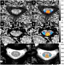
          Anatomical magnetic resonance imaging (MRI) is well established in the literature as the key imaging tool to identify spinal cord compression, disc herniation/bulging, and inbuckling of the ligamentum flavum, thus facilitating surgical planning, while radiographs and computed tomography (CT) provide complimentary information. Electrophysiology techniques are primarily used to rule out competing diagnoses. However, signal change and measures of cord compression on conventional MRI have limited utility to characterize the degree of tissue injury, which may be helpful for diagnosis, prognostication, and repeated assessments to identify deterioration. Early translational studies of quantitative imaging and electrophysiology techniques show potential of these methods to more accurately reflect changes in spinal cord microstructure and function.

          Conclusion

          Currently, clinical management of DCM relies heavily on anatomical MRI, with additional contributions from radiographs, CT, and electrophysiology. Novel quantitative assessments of microstructure, perfusion, and function have the potential to transform clinical practice, but require robust validation, automation, and standardization prior to uptake.

          Related collections

          Most cited references147

          • Record: found
          • Abstract: found
          • Article: not found

          Adjacent segment degeneration and adjacent segment disease: the consequences of spinal fusion?

          Given the number of spinal fusions performed annually, concerns have mounted over the potential for adjacent segment degeneration (radiographic changes of degeneration at levels adjacent to a spinal fusion) and adjacent segment disease (development of new symptoms correlating with adjacent segment degeneration). This article reviews documented evidence on adjacent segment degeneration and disease as it relates to cervical and lumbar arthrodesis. There appears to be an incidence of adjacent segment degeneration and disease after arthrodesis that may be related to natural degeneration or the adjacent fusion. It remains to be seen whether restoration of motion with disc arthroplasty will alter the rate of adjacent segment degeneration or disease.
            Bookmark
            • Record: found
            • Abstract: found
            • Article: not found

            Diaschisis: past, present, future.

            After a century of false hopes, recent studies have placed the concept of diaschisis at the centre of the understanding of brain function. Originally, the term 'diaschisis' was coined by von Monakow in 1914 to describe the neurophysiological changes that occur distant to a focal brain lesion. In the following decades, this concept triggered widespread clinical interest in an attempt to describe symptoms and signs that the lesion could not fully explain. However, the first imaging studies, in the late 1970s, only partially confirmed the clinical significance of diaschisis. Focal cortical areas of diaschisis (i.e. focal diaschisis) contributed to the clinical deficits after subcortical but only rarely after cortical lesions. For this reason, the concept of diaschisis progressively disappeared from the mainstream of research in clinical neurosciences. Recent evidence has unexpectedly revitalized the notion. The development of new imaging techniques allows a better understanding of the complexity of brain organization. It is now possible to reliably investigate a new type of diaschisis defined as the changes of structural and functional connectivity between brain areas distant to the lesion (i.e. connectional diaschisis). As opposed to focal diaschisis, connectional diaschisis, focusing on determined networks, seems to relate more consistently to the clinical findings. This is particularly true after stroke in the motor and attentional networks. Furthermore, normalization of remote connectivity changes in these networks relates to a better recovery. In the future, to investigate the clinical role of diaschisis, a systematic approach has to be considered. First, emerging imaging and electrophysiological techniques should be used to precisely map and selectively model brain lesions in human and animals studies. Second, the concept of diaschisis must be applied to determine the impact of a focal lesion on new representations of the complexity of brain organization. As an example, the evaluation of remote changes in the structure of the connectome has so far mainly been tested by modelization of focal brain lesions. These changes could now be assessed in patients suffering from focal brain lesions (i.e. connectomal diaschisis). Finally, and of major significance, focal and non-focal neurophysiological changes distant to the lesion should be the target of therapeutic strategies. Neuromodulation using transcranial magnetic stimulation is one of the most promising techniques. It is when this last step will be successful that the concept of diaschisis will gain all the clinical respectability that could not be obtained in decades of research. © The Author (2014). Published by Oxford University Press on behalf of the Guarantors of Brain. All rights reserved. For Permissions, please email: journals.permissions@oup.com.
              Bookmark
              • Record: found
              • Abstract: found
              • Article: not found

              Degenerative Cervical Myelopathy: Epidemiology, Genetics, and Pathogenesis.

              Review.
                Bookmark

                Author and article information

                Journal
                Global Spine J
                Global Spine J
                spgsj
                GSJ
                Global Spine Journal
                SAGE Publications (Sage CA: Los Angeles, CA )
                2192-5682
                2192-5690
                19 November 2021
                February 2022
                : 12
                : 1 Suppl , Special Issue: Transforming Care for Degenerative Cervical Myelopathy: The Top Ten Priorities. Findings in Context From AO Spine RECODE-DCM, an International and Multi-Disciplinary Collaboration
                : 130S-146S
                Affiliations
                [1 ]Department of Neurological Surgery, Ringgold 8789, universityUniversity of California Davis; , Davis, CA, USA
                [2 ]Department of Neurology, Ringgold 5894, universityNew York University; , Langone Health, Graduate Medical Education, New York, NY, USA
                [3 ]Division of Neurosurgery, Geneva University Hospitals, Ringgold 27230, universityUniversity of Geneva; , Geneva, Switzerland
                [4 ]universityUniversity Spine Center; , Ringgold 31031, universityBalgrist University Hospital; , Zurich, Switzerland
                [5 ]Department of Neurosurgery, Sina Trauma and Surgery Research Center, Ringgold 48439, universityTehran University of Medical Sciences; , Tehran, Iran
                [6 ]Division of Neurosurgery, Department of Surgery, Ringgold 7938, universityUniversity of Toronto; , Toronto, ON, Canada
                [7 ]Vancouver Spine Surgery Institute, Department of Orthopedics, universityThe University of British Columbia; , Vancouver, BC, Canada
                [8 ]Department of Neurological Surgery, Ringgold 6529, universityThomas Jefferson University; , Philadelphia, PA, USA
                [9 ]Department of Neurosurgery, Ringgold 2152, universityUniversity of Cambridge; , Cambridge, UK
                [10 ]Department of Neurosurgery and The Miami Project to Cure Paralysis, The Miller School of Medicine, Ringgold 12235, universityUniversity of Miami; , Miami, FL, USA
                [11 ]Department of Neurosurgery, Ringgold 1479, universityUniversity of Maryland; , Baltimore, MD, USA
                [12 ]Department of Neurosurgery, Ringgold 5506, universityMedical College of Wisconsin; , Wauwatosa, WI, USA
                Author notes
                [*]Allan R. Martin, Department of Neurological Surgery, University of California Davis, Davis, CA, USA. Email: armartin@ 123456ucdavis.edu
                Author information
                https://orcid.org/0000-0002-4965-3059
                https://orcid.org/0000-0001-5965-0305
                https://orcid.org/0000-0002-5722-6364
                https://orcid.org/0000-0003-0591-5069
                Article
                10.1177_21925682211057484
                10.1177/21925682211057484
                8859711
                34797993
                c245250a-f6b4-4d72-96f8-0ab67dfe4529
                © The Author(s) 2021

                This article is distributed under the terms of the Creative Commons Attribution-NonCommercial-NoDerivs 4.0 License ( https://creativecommons.org/licenses/by-nc-nd/4.0/) which permits non-commercial use, reproduction and distribution of the work as published without adaptation or alteration, without further permission provided the original work is attributed as specified on the SAGE and Open Access pages ( https://us.sagepub.com/en-us/nam/open-access-at-sage).

                History
                Categories
                Special Issue Articles
                Custom metadata
                ts10

                cervical myelopathy,imaging,diagnosis,electrophysiology,spinal cord compression,magnetic resonance,neural damage,microstructure,assessment

                Comments

                Comment on this article