1
views
0
recommends
+1 Recommend
0 collections
    0
    shares
      • Record: found
      • Abstract: found
      • Article: found
      Is Open Access

      Increased Prefrontal Activation During Verbal Fluency Task After Repetitive Transcranial Magnetic Stimulation Treatment in Depression: A Functional Near-Infrared Spectroscopy Study

      research-article

      Read this article at

      Bookmark
          There is no author summary for this article yet. Authors can add summaries to their articles on ScienceOpen to make them more accessible to a non-specialist audience.

          Abstract

          Background

          Previous studies have shown the clinical effect of 2 Hz repetitive transcranial magnetic stimulation (rTMS) for depression; however, its underlying neural mechanisms are poorly understood. The aim of this study was to examine the effects of rTMS on the activity of the prefrontal cortex in patients with depression, using functional near-infrared spectroscopy (fNIRS).

          Methods

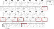
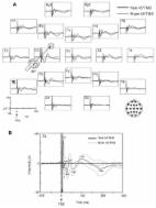
          Forty patients with major depressive disorder (MDD) and 40 healthy controls were enrolled in this study. Patients underwent 4 weeks of 2 Hz TMS delivered to the right dorsolateral prefrontal cortex (DLPFC). fNIRS was used to measure the changes in the concentration of oxygenated hemoglobin ([oxy-Hb]) in the prefrontal cortex during a verbal fluency task (VFT) in depressed patients before and after rTMS treatment. The severity of depression was assessed using the Hamilton Rating Scale for Depression-24 item (HAMD-24).

          Results

          Prior to rTMS, depressed patients exhibited significantly smaller [oxy-Hb] values in the bilateral prefrontal cortex during the VFT compared with the healthy controls. After 4 weeks of 2 Hz right DLPFC rTMS treatment, increased [oxy-Hb] values in the bilateral frontopolar prefrontal cortex (FPPFC), ventrolateral prefrontal cortex (VLPFC) and left DLPFC during the VFT were observed in depressed patients. The increased [oxy-Hb] values from baseline to post-treatment in the right VLPFC in depressed patients were positively related to the reduction of HAMD score following rTMS.

          Conclusion

          These findings suggest that the function of the prefrontal cortex in depressed patients was impaired and could be recovered by 2 Hz rTMS. The fNIRS-measured prefrontal activation during a cognitive task is a potential biomarker for monitoring depressed patients’ treatment response to rTMS.

          Related collections

          Most cited references70

          • Record: found
          • Abstract: found
          • Article: not found

          Acute and Longer-Term Outcomes in Depressed Outpatients Requiring One or Several Treatment Steps: A STAR*D Report

          This report describes the participants and compares the acute and longer-term treatment outcomes associated with each of four successive steps in the Sequenced Treatment Alternatives to Relieve Depression (STAR*D) trial.
            Bookmark
            • Record: found
            • Abstract: found
            • Article: found
            Is Open Access

            Non-invasive electrical and magnetic stimulation of the brain, spinal cord, roots and peripheral nerves: Basic principles and procedures for routine clinical and research application. An updated report from an I.F.C.N. Committee

            These guidelines provide an up-date of previous IFCN report on “Non-invasive electrical and magnetic stimulation of the brain, spinal cord and roots: basic principles and procedures for routine clinical application” (Rossini et al., 1994). A new Committee, composed of international experts, some of whom were in the panel of the 1994 “Report”, was selected to produce a current state-of-the-art review of non-invasive stimulation both for clinical application and research in neuroscience. Since 1994, the international scientific community has seen a rapid increase in non-invasive brain stimulation in studying cognition, brain–behavior relationship and pathophysiology of various neurologic and psychiatric disorders. New paradigms of stimulation and new techniques have been developed. Furthermore, a large number of studies and clinical trials have demonstrated potential therapeutic applications of non-invasive brain stimulation, especially for TMS. Recent guidelines can be found in the literature covering specific aspects of non-invasive brain stimulation, such as safety (Rossi et al., 2009), methodology (Groppa et al., 2012) and therapeutic applications (Lefaucheur et al., 2014). This up-dated review covers theoretical, physiological and practical aspects of non-invasive stimulation of brain, spinal cord, nerve roots and peripheral nerves in the light of more updated knowledge, and include some recent extensions and developments.
              Bookmark
              • Record: found
              • Abstract: found
              • Article: not found

              HomER: a review of time-series analysis methods for near-infrared spectroscopy of the brain.

              Near-infrared spectroscopy (NIRS) is a noninvasive neuroimaging tool for studying evoked hemodynamic changes within the brain. By this technique, changes in the optical absorption of light are recorded over time and are used to estimate the functionally evoked changes in cerebral oxyhemoglobin and deoxyhemoglobin concentrations that result from local cerebral vascular and oxygen metabolic effects during brain activity. Over the past three decades this technology has continued to grow, and today NIRS studies have found many niche applications in the fields of psychology, physiology, and cerebral pathology. The growing popularity of this technique is in part associated with a lower cost and increased portability of NIRS equipment when compared with other imaging modalities, such as functional magnetic resonance imaging and positron emission tomography. With this increasing number of applications, new techniques for the processing, analysis, and interpretation of NIRS data are continually being developed. We review some of the time-series and functional analysis techniques that are currently used in NIRS studies, we describe the practical implementation of various signal processing techniques for removing physiological, instrumental, and motion-artifact noise from optical data, and we discuss the unique aspects of NIRS analysis in comparison with other brain imaging modalities. These methods are described within the context of the MATLAB-based graphical user interface program, HomER, which we have developed and distributed to facilitate the processing of optical functional brain data.
                Bookmark

                Author and article information

                Contributors
                Journal
                Front Psychiatry
                Front Psychiatry
                Front. Psychiatry
                Frontiers in Psychiatry
                Frontiers Media S.A.
                1664-0640
                04 April 2022
                2022
                : 13
                : 876136
                Affiliations
                [1] 1Mental Health Center, West China Hospital, West China School of Medicine, Sichuan University , Chengdu, China
                [2] 2Department of Rehabilitation Sciences, The Hong Kong Polytechnic University, Kowloon , Hong Kong SAR, China
                [3] 3Department of Rehabilitation Medicine, The Seventh Affiliated Hospital, Sun Yat-sen University , Shenzhen, China
                [4] 4Guangdong Engineering and Technology Research Center for Rehabilitation Medicine and Translation , Guangzhou, China
                Author notes

                Edited by: Po-Han Chou, China Medical University Hospital, Taiwan

                Reviewed by: Che-sheng Chu, Kaohsiung Veterans General Hospital, Taiwan; Hsin-An Chang, Tri-Service General Hospital, Taiwan

                *Correspondence: Zhong Zheng, zhengzhong@ 123456wchscu.cn

                These authors have contributed equally to this work

                This article was submitted to Neuroimaging and Stimulation, a section of the journal Frontiers in Psychiatry

                Article
                10.3389/fpsyt.2022.876136
                9013767
                35444573
                c44e7e37-243a-49ef-8b47-e69c979d19e1
                Copyright © 2022 Huang, Zhang, Zhang, Wang and Zheng.

                This is an open-access article distributed under the terms of the Creative Commons Attribution License (CC BY). The use, distribution or reproduction in other forums is permitted, provided the original author(s) and the copyright owner(s) are credited and that the original publication in this journal is cited, in accordance with accepted academic practice. No use, distribution or reproduction is permitted which does not comply with these terms.

                History
                : 15 February 2022
                : 09 March 2022
                Page count
                Figures: 5, Tables: 1, Equations: 0, References: 70, Pages: 10, Words: 7433
                Funding
                Funded by: National Natural Science Foundation of China, doi 10.13039/501100001809;
                Funded by: Sichuan Province Science and Technology Support Program, doi 10.13039/100012542;
                Categories
                Psychiatry
                Original Research

                Clinical Psychology & Psychiatry
                major depressive disorder,repetitive transcranial magnetic stimulation,prefrontal cortex,functional near-infrared spectroscopy,verbal fluency task

                Comments

                Comment on this article