2
views
0
recommends
+1 Recommend
0 collections
    0
    shares
      • Record: found
      • Abstract: found
      • Article: found
      Is Open Access

      Two complementary approaches for efficient isolation of Sertoli cells for transcriptomic analysis

      methods-article

      Read this article at

      Bookmark
          There is no author summary for this article yet. Authors can add summaries to their articles on ScienceOpen to make them more accessible to a non-specialist audience.

          Abstract

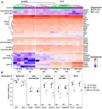
          Sertoli cells (SCs) are the only somatic cells that reside in seminiferous tubules of testis. They directly interact with and support the development of germ cells, thus have an indispensable role in the process of spermatogenesis. SCs first appear in a proliferative state and then, with the initiation of the first wave of spermatogenesis, progress to a mature “nurturing” state which supports lifelong continuous sperm production. During this development, the SC transcriptome must adapt rapidly as obstacles in SC maturation often result in deficiencies in male fertility. Due to its importance in spermatogenesis, a reliable, rapid, and precise method for the isolation of high purity, viable and unadulterated SC has been largely missing. We have developed an improved method for the preparation of a testicular single cell suspension comprised of two alternative protocols to separate SCs from the rest of the testicular cells by FACS. The first sorting scheme is based on their co-expression of surface specific markers, FSHr and Occludin-1, while the second focuses on the co-staining of SCs with FSHr-specific antibody and Hoechst 33342, which discriminates DNA content of testicular cells. The entire procedure can be completed in less than 3 h which permits the analysis of the development-related transcriptional profile of these cells. Notably, our comparative study showed that this method resulted in a SC transcriptome that is largely comparable to SCs which were briskly isolated due to their cell-specific expression of fluorescent protein. Interestingly, we also show that SCs sorted as FSHr +Occludin + cells contained a tangible portion of transcripts from all types of testicular germ cells. Sorting of SCs according to their 2C DNA content significantly reduced the presence of these transcripts, thus seems to be the most suitable approach for accurate determination of the SC transcriptome. We believe that these novel approaches for the isolation of SCs will assist researchers in the elucidation of their function as well as their role in spermatogenesis and disorders related to male infertility.

          Related collections

          Most cited references54

          • Record: found
          • Abstract: found
          • Article: not found

          A new mathematical model for relative quantification in real-time RT-PCR.

          M. Pfaffl (2001)
          Use of the real-time polymerase chain reaction (PCR) to amplify cDNA products reverse transcribed from mRNA is on the way to becoming a routine tool in molecular biology to study low abundance gene expression. Real-time PCR is easy to perform, provides the necessary accuracy and produces reliable as well as rapid quantification results. But accurate quantification of nucleic acids requires a reproducible methodology and an adequate mathematical model for data analysis. This study enters into the particular topics of the relative quantification in real-time RT-PCR of a target gene transcript in comparison to a reference gene transcript. Therefore, a new mathematical model is presented. The relative expression ratio is calculated only from the real-time PCR efficiencies and the crossing point deviation of an unknown sample versus a control. This model needs no calibration curve. Control levels were included in the model to standardise each reaction run with respect to RNA integrity, sample loading and inter-PCR variations. High accuracy and reproducibility (<2.5% variation) were reached in LightCycler PCR using the established mathematical model.
            Bookmark
            • Record: found
            • Abstract: found
            • Article: not found

            Spermatogenic cells of the prepuberal mouse: isolation and morphological characterization

            A procedure is described which permits the isolation from the prepuberal mouse testis of highly purified populations of primitive type A spermatogonia, type A spermatogonia, type B spermatogonia, preleptotene primary spermatocytes, leptotene and zygotene primary spermatocytes, pachytene primary spermatocytes and Sertoli cells. The successful isolation of these prepuberal cell types was accomplished by: (a) defining distinctive morphological characteristics of the cells, (b) determining the temporal appearance of spermatogenic cells during prepuberal development, (c) isolating purified seminiferous cords, after dissociation of the testis with collagenase, (d) separating the trypsin-dispersed seminiferous cells by sedimentation velocity at unit gravity, and (e) assessing the identity and purity of the isolated cell types by microscopy. The seminiferous epithelium from day 6 animals contains only primitive type A spermatogonia and Sertoli cells. Type A and type B spermatogonia are present by day 8. At day 10, meiotic prophase is initiated, with the germ cells reaching the early and late pachytene stages by 14 and 18, respectively. Secondary spermatocytes and haploid spermatids appear throughout this developmental period. The purity and optimum day for the recovery of specific cell types are as follows: day 6, Sertoli cells (purity>99 percent) and primitive type A spermatogonia (90 percent); day 8, type A spermatogonia (91 percent) and type B spermatogonia (76 percent); day 18, preleptotene spermatocytes (93 percent), leptotene/zygotene spermatocytes (52 percent), and pachytene spermatocytes (89 percent), leptotene/zygotene spermatocytes (52 percent), and pachytene spermatocytes (89 percent).
              Bookmark
              • Record: found
              • Abstract: found
              • Article: not found

              The Sertoli cell: one hundred fifty years of beauty and plasticity.

              It has been one and a half centuries since Enrico Sertoli published the seminal discovery of the testicular 'nurse cell', not only a key cell in the testis, but indeed one of the most amazing cells in the vertebrate body. In this review, we begin by examining the three phases of morphological research that have occurred in the study of Sertoli cells, because microscopic anatomy was essentially the only scientific discipline available for about the first 75 years after the discovery. Biochemistry and molecular biology then changed all of biological sciences, including our understanding of the functions of Sertoli cells. Immunology and stem cell biology were not even topics of science in 1865, but they have now become major issues in our appreciation of Sertoli cell's role in spermatogenesis. We end with the universal importance and plasticity of function by comparing Sertoli cells in fish, amphibians, and mammals. In these various classes of vertebrates, Sertoli cells have quite different modes of proliferation and epithelial maintenance, cystic vs. tubular formation, yet accomplish essentially the same function but in strikingly different ways.
                Bookmark

                Author and article information

                Contributors
                Journal
                Front Cell Dev Biol
                Front Cell Dev Biol
                Front. Cell Dev. Biol.
                Frontiers in Cell and Developmental Biology
                Frontiers Media S.A.
                2296-634X
                06 September 2022
                2022
                : 10
                : 972017
                Affiliations
                [1] 1 Laboratory of Immunobiology , Institute of Molecular Genetics of the Czech Academy of Sciences , Prague, Czechia
                [2] 2 Laboratory of Genomics and Bioinformatics , Institute of Molecular Genetics of the Czech Academy of Sciences , Prague, Czechia
                Author notes

                Edited by: Ciler Celik-Ozenci, Koç University, Turkey

                Reviewed by: Isabelle Stévant, Bar-Ilan University, Israel

                Sevil Cayli, Ankara Yıldırım Beyazıt University, Turkey

                *Correspondence: Dominik Filipp, dominik.filipp@ 123456img.cas.cz

                This article was submitted to Molecular and Cellular Reproduction, a section of the journal Frontiers in Cell and Developmental Biology

                Article
                972017
                10.3389/fcell.2022.972017
                9495933
                c4981d46-1ea5-44a5-96db-42bd1d06e042
                Copyright © 2022 Petrusová, Manning, Kubovčiak, Kolář and Filipp.

                This is an open-access article distributed under the terms of the Creative Commons Attribution License (CC BY). The use, distribution or reproduction in other forums is permitted, provided the original author(s) and the copyright owner(s) are credited and that the original publication in this journal is cited, in accordance with accepted academic practice. No use, distribution or reproduction is permitted which does not comply with these terms.

                History
                : 17 June 2022
                : 12 August 2022
                Funding
                Funded by: Grantová Agentura České Republiky , doi 10.13039/501100001824;
                Categories
                Cell and Developmental Biology
                Methods

                sertoli cell,flow cytometry,fshr,occludin,spermatogenesis
                sertoli cell, flow cytometry, fshr, occludin, spermatogenesis

                Comments

                Comment on this article