1
views
0
recommends
+1 Recommend
0 collections
    0
    shares
      • Record: found
      • Abstract: found
      • Article: found
      Is Open Access

      Histological Correlates of Neuroanatomical Changes in a Rat Model of Levodopa-Induced Dyskinesia Based on Voxel-Based Morphometry

      research-article

      Read this article at

      Bookmark
          There is no author summary for this article yet. Authors can add summaries to their articles on ScienceOpen to make them more accessible to a non-specialist audience.

          Abstract

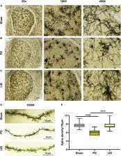
          Long-term therapy with levodopa (L-DOPA) in patients with Parkinson’s disease (PD) often triggers motor complications termed as L-DOPA-induced dyskinesia (LID). However, few studies have explored the pathogenesis of LID from the perspective of neuroanatomy. This study aimed to investigate macroscopic structural changes in a rat model of LID and the underlying histological mechanisms. First, we established the hemiparkinsonism rat model through stereotaxic injection of 6-hydroxydopamine (6-OHDA) into the right medial forebrain bundle, followed by administration of saline (PD) or L-DOPA to induce LID. Magnetic resonance imaging (MRI) and behavioral evaluations were performed at different time points. Histological analysis was conducted to assess the correlations between MRI signal changes and cellular contributors. Voxel-based morphometry (VBM) analysis revealed progressive bilateral volume reduction in the cortical and subcortical areas in PD rats compared with the sham rats. These changes were partially reversed by chronic L-DOPA administration; moreover, there was a significant volume increase mainly in the dorsolateral striatum, substantia nigra, and piriform cortex of the lesioned side compared with that of PD rats. At the striatal cellular level, glial fibrillary acidic protein-positive (GFAP+) astrocytes were significantly increased in the lesioned dorsolateral striatum of PD rats compared with the intact side and the sham group. Prolonged L-DOPA treatment further increased GFAP levels. Neither 6-OHDA damage nor L-DOPA treatment influenced the striatal expression of vascular endothelial growth factor (VEGF). Additionally, there was a considerable increase in synapse-associated proteins (SYP, PSD95, and SAP97) in the lesioned striatum of LID rats relative to the PD rats. Golgi-Cox staining analysis of the dendritic spine morphology revealed an increased density of dendritic spines after chronic L-DOPA treatment. Taken together, our findings suggest that striatal volume changes in LID rats involve astrocyte activation, enrichment of synaptic ultrastructure and signaling proteins in the ipsilateral striatum. Meanwhile, the data highlight the enormous potential of structural MRI, especially VBM analysis, in determining the morphological phenotype of rodent models of LID.

          Related collections

          Most cited references102

          • Record: found
          • Abstract: found
          • Article: not found

          Voxel-based morphometry--the methods.

          At its simplest, voxel-based morphometry (VBM) involves a voxel-wise comparison of the local concentration of gray matter between two groups of subjects. The procedure is relatively straightforward and involves spatially normalizing high-resolution images from all the subjects in the study into the same stereotactic space. This is followed by segmenting the gray matter from the spatially normalized images and smoothing the gray-matter segments. Voxel-wise parametric statistical tests which compare the smoothed gray-matter images from the two groups are performed. Corrections for multiple comparisons are made using the theory of Gaussian random fields. This paper describes the steps involved in VBM, with particular emphasis on segmenting gray matter from MR images with nonuniformity artifact. We provide evaluations of the assumptions that underpin the method, including the accuracy of the segmentation and the assumptions made about the statistical distribution of the data. Copyright 2000 Academic Press.
            Bookmark
            • Record: found
            • Abstract: found
            • Article: not found

            Tripartite synapses: astrocytes process and control synaptic information.

            The term 'tripartite synapse' refers to a concept in synaptic physiology based on the demonstration of the existence of bidirectional communication between astrocytes and neurons. Consistent with this concept, in addition to the classic 'bipartite' information flow between the pre- and postsynaptic neurons, astrocytes exchange information with the synaptic neuronal elements, responding to synaptic activity and, in turn, regulating synaptic transmission. Because recent evidence has demonstrated that astrocytes integrate and process synaptic information and control synaptic transmission and plasticity, astrocytes, being active partners in synaptic function, are cellular elements involved in the processing, transfer and storage of information by the nervous system. Consequently, in contrast to the classically accepted paradigm that brain function results exclusively from neuronal activity, there is an emerging view, which we review herein, in which brain function actually arises from the coordinated activity of a network comprising both neurons and glia.
              Bookmark
              • Record: found
              • Abstract: found
              • Article: not found

              Mechanisms of angiogenesis and arteriogenesis.

              Endothelial and smooth muscle cells interact with each other to form new blood vessels. In this review, the cellular and molecular mechanisms underlying the formation of endothelium-lined channels (angiogenesis) and their maturation via recruitment of smooth muscle cells (arteriogenesis) during physiological and pathological conditions are summarized, alongside with possible therapeutic applications.
                Bookmark

                Author and article information

                Contributors
                Journal
                Front Aging Neurosci
                Front Aging Neurosci
                Front. Aging Neurosci.
                Frontiers in Aging Neuroscience
                Frontiers Media S.A.
                1663-4365
                28 October 2021
                2021
                : 13
                : 759934
                Affiliations
                [1] 1Department of Neurology, Union Hospital, Tongji Medical College, Huazhong University of Science and Technology , Wuhan, China
                [2] 2State Key Laboratory of Magnetic Resonance and Atomic and Molecular Physics, Innovation Academy for Precision Measurement Science and Technology, National Center for Magnetic Resonance in Wuhan, Chinese Academy of Sciences , Wuhan, China
                [3] 3University of Chinese Academy of Sciences , Beijing, China
                Author notes

                Edited by: Francesco Napolitano, Università degli Studi di Napoli Federico II, Italy

                Reviewed by: Alessandra Bonito-Oliva, The Rockefeller University, United States; Veronica Ghiglieri, Università Telematica San Raffaele Roma, Italy

                *Correspondence: Hao Lei, leihao@ 123456wipm.ac.cn

                These authors have contributed equally to this work

                Article
                10.3389/fnagi.2021.759934
                8581620
                34776935
                c65c1b92-0006-4a5a-94fd-86a4ba908249
                Copyright © 2021 Zhang, Chen, Wu, Zeng, Yuan, Cheng, Yang, Wang, Yang, Xu, Lei, Cao and Xu.

                This is an open-access article distributed under the terms of the Creative Commons Attribution License (CC BY). The use, distribution or reproduction in other forums is permitted, provided the original author(s) and the copyright owner(s) are credited and that the original publication in this journal is cited, in accordance with accepted academic practice. No use, distribution or reproduction is permitted which does not comply with these terms.

                History
                : 17 August 2021
                : 07 October 2021
                Page count
                Figures: 7, Tables: 0, Equations: 0, References: 102, Pages: 17, Words: 13151
                Categories
                Aging Neuroscience
                Original Research

                Neurosciences
                parkinson’s disease,l-dopa-induced dyskinesia,magnetic resonance imaging,voxel-based morphometry,striatum,astrocyte,synaptic plasticity,microvasculature

                Comments

                Comment on this article