31
views
0
recommends
+1 Recommend
0 collections
    0
    shares
      • Record: found
      • Abstract: found
      • Article: not found

      Compact Polyelectrolyte Complexes: “Saloplastic” Candidates for Biomaterials

      research-article
      ,
      Biomacromolecules
      American Chemical Society

      Read this article at

      ScienceOpenPublisherPMC
      Bookmark
          There is no author summary for this article yet. Authors can add summaries to their articles on ScienceOpen to make them more accessible to a non-specialist audience.

          Abstract

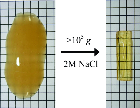
          Precipitates of polyelectrolyte complexes were transformed into rugged shapes suitable for bioimplants by ultracentrifugation in the presence of high salt concentration. Salt ions dope the complex, creating a softer material with viscous fluid-like properties. Complexes that were compacted under the centrifugal field (CoPECs) were made from poly(diallyldimethyl ammonium), PDADMA, as polycation, and poly(styrene sulfonate), PSS, or poly(methacrylic acid), PMAA, as polyanion. Dynamic mechanical testing revealed a rubbery plateau at lower frequencies for PSS/PDADMA with moduli that decreased with increasing salt concentration, as internal ion pair cross-links were broken. CoPECs had significantly lower modulii compared to similar polyelectrolyte complexes prepared by the “multilayering” method. The difference in mechanical properties was ascribed to higher water content (located in micropores) for the former and, more importantly, to their nonstoichiometric polymer composition. The modulus of PMAA/PDADMA CoPECs, under physiological conditions, demonstrated dynamic mechanical properties that were close to those of the nucleus pulposus in an intervertebral disk.

          Related collections

          Most cited references32

          • Record: found
          • Abstract: found
          • Article: not found

          Versatile Photocatalytic Systems for H2 Generation in Water Based on an Efficient DuBois-Type Nickel Catalyst

          The generation of renewable H2 through an efficient photochemical route requires photoinduced electron transfer (ET) from a light harvester to an efficient electrocatalyst in water. Here, we report on a molecular H2 evolution catalyst (NiP) with a DuBois-type [Ni(P2 R′N2 R″)2]2+ core (P2 R′N2 R″ = bis(1,5-R′-diphospha-3,7-R″-diazacyclooctane), which contains an outer coordination sphere with phosphonic acid groups. The latter functionality allows for good solubility in water and immobilization on metal oxide semiconductors. Electrochemical studies confirm that NiP is a highly active electrocatalyst in aqueous electrolyte solution (overpotential of approximately 200 mV at pH 4.5 with a Faradaic yield of 85 ± 4%). Photocatalytic experiments and investigations on the ET kinetics were carried out in combination with a phosphonated Ru(II) tris(bipyridine) dye (RuP) in homogeneous and heterogeneous environments. Time-resolved luminescence and transient absorption spectroscopy studies confirmed that directed ET from RuP to NiP occurs efficiently in all systems on the nano- to microsecond time scale, through three distinct routes: reductive quenching of RuP in solution or on the surface of ZrO2 (“on particle” system) or oxidative quenching of RuP when the compounds were immobilized on TiO2 (“through particle” system). Our studies show that NiP can be used in a purely aqueous solution and on a semiconductor surface with a high degree of versatility. A high TOF of 460 ± 60 h–1 with a TON of 723 ± 171 for photocatalytic H2 generation with a molecular Ni catalyst in water and a photon-to-H2 quantum yield of approximately 10% were achieved for the homogeneous system.
            Bookmark
            • Record: found
            • Abstract: found
            • Article: not found

            The chondrocyte, architect of cartilage. Biomechanics, structure, function and molecular biology of cartilage matrix macromolecules.

            Chondrocytes are specialised cells which produce and maintain the extracellular matrix of cartilage, a tissue that is resilient and pliant. In vivo, it has to withstand very high compressive loads, and that is explicable in terms of the physico-chemical properties of cartilage-specific macromolecules and with the movement of water and ions within the matrix. The functions of the cartilage-specific collagens, aggrecan (a hydrophilic proteoglycan) and hyaluronan are discussed within this context. The structures of cartilage collagens and proteoglycans and their genes are known and a number of informative mutations have been identified. In particular, collagen fibrillogenesis is a complex process which can be altered by mutations whose effects fit what is known about collagen molecular structural functions. In other instances, mutations have indicated new functions for particular molecular domains. As cartilage provides the template for the developing skeleton, mutations in genes for cartilage-specific proteins often produce developmental abnormalities. The search for mutations amongst such genes in heritable disorders is being actively pursued by many groups, although mutation and phenotype are not always well correlated, probably because of compensatory mechanisms. The special nature of the chondrocyte is stressed in connection with its cell involvement in osteoarthritis, the most widespread disease of diarthrodial joints.
              Bookmark
              • Record: found
              • Abstract: found
              • Article: not found

              Scaffold design and fabrication technologies for engineering tissues--state of the art and future perspectives.

              Today, tissue engineers are attempting to engineer virtually every human tissue. Potential tissue-engineered products include cartilage, bone, heart valves, nerves, muscle, bladder, liver, etc. Tissue engineering techniques generally require the use of a porous scaffold, which serves as a three-dimensional template for initial cell attachment and subsequent tissue formation both in vitro and in vivo. The scaffold provides the necessary support for cells to attach, proliferate, and maintain their differentiated function. Its architecture defines the ultimate shape of the new grown soft or hard tissue. In the early days of tissue engineering, clinically established materials such as collagen and polyglycolide were primarily considered as the material of choice for scaffolds. The challenge for more advanced scaffold systems is to arrange cells/tissue in an appropriate 3D configuration and present molecular signals in an appropriate spatial and temporal fashion so that the individual cells will grow and form the desired tissue structures--and do so in a way that can be carried out reproducibly, economically, and on a large scale. This paper is not intended to provide a general review of tissue engineering, but specifically concentrate on the design and processing of synthetic polymeric scaffolds. The material properties and design requirements are discussed. An overview of the various fabrication techniques of scaffolds is presented, beginning with the basic and conventional techniques to the more recent, novel methods that combine both scaffold design and fabrication capabilities.
                Bookmark

                Author and article information

                Journal
                Biomacromolecules
                bm
                bomaf6
                Biomacromolecules
                American Chemical Society
                1525-7797
                1526-4602
                19 October 2009
                09 November 2009
                : 10
                : 11
                : 2968-2975
                Affiliations
                Department of Chemistry and Biochemistry, The Florida State University, Tallahassee, Florida 32306
                Author notes
                [* ] To whom correspondence should be addressed. E-mail: schlen@ 123456chem.fsu.edu .
                [†]

                Current address: ICO Polymers, 77130 Montereau Fault Yonne, France.

                Article
                10.1021/bm900373c
                2774624
                19835412
                c79c0d07-dc1a-4b61-a35f-cd29a65202a4
                Copyright © 2009 American Chemical Society

                This is an open-access article distributed under the ACS AuthorChoice Terms & Conditions. Any use of this article, must conform to the terms of that license which are available at http://pubs.acs.org.

                History
                : 2 April 2009
                : 9 July 2009
                : 19 October 2009
                : 09 November 2009
                Funding
                National Institutes of Health, United States
                Categories
                Article
                Custom metadata
                bm900373c
                bm-2009-00373c
                40.75

                Biochemistry
                Biochemistry

                Comments

                Comment on this article