20
views
0
recommends
+1 Recommend
0 collections
    0
    shares
      • Record: found
      • Abstract: found
      • Article: found
      Is Open Access

      Fibrous dysplasia of the anterior mandible: A rare case report

      case-report

      Read this article at

      Bookmark
          There is no author summary for this article yet. Authors can add summaries to their articles on ScienceOpen to make them more accessible to a non-specialist audience.

          Abstract

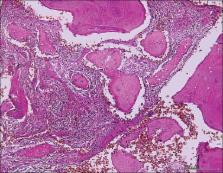
          Fibrous dysplasia (FD) is a rare bony disorder in which normal bone is replaced by abnormal fibro-osseous tissue. It often involves the long bones, craniofacial bones, ribs, and pelvis. Approximately 30% of monostotic FD (MFD) lesions are found in the cranial or facial bones. In general, FD is found in teenagers, and it usually becomes static after adulthood. FD involves the maxilla almost two times more often than the mandible. It frequently appears in the posterior region of the jaw bone and is usually unilateral. Here, we present an unusual case of symptomatic MFD affecting the anterior region of the mandible in a 43-year-old female with the clinical, radiographical, and histopathological features. The clinical examination showed both the labial and lingual bone expansion in the anterior mandible. The radiographic examination revealed a lesion with both radiopaque and radiolucent features showing a “ground-glass” appearance. The diagnosis was obtained after confirmatory intrabony biopsy with the histopathological examination, and it was diagnosed with benign FD. The patient preferred regular follow-up of MFD after discussion. During the regular follow-up, MFD lesion showed no obvious signs of progression or malignancy features.

          Related collections

          Most cited references9

          • Record: found
          • Abstract: found
          • Article: not found

          Fibrous dysplasia. Pathophysiology, evaluation, and treatment.

          Fibrous dysplasia is a common benign skeletal lesion that may involve one bone (monostotic) or multiple bones (polyostotic) and occurs throughout the skeleton with a predilection for the long bones, ribs, and craniofacial bones. The etiology of fibrous dysplasia has been linked to an activating mutation in the gene that encodes the alpha subunit of stimulatory G protein (G(s)alpha) located at 20q13.2-13.3. Most lesions are monostotic, asymptomatic, and identified incidentally and can be treated with clinical observation and patient education. Bisphosphonate therapy may help to improve function, decrease pain, and lower fracture risk in appropriately selected patients with fibrous dysplasia. Surgery is indicated for confirmatory biopsy, correction of deformity, prevention of pathologic fracture, and/or eradication of symptomatic lesions. The use of cortical grafts is preferred over cancellous grafts or bone-graft substitutes because of the superior physical qualities of remodeled cortical bone.
            Bookmark
            • Record: found
            • Abstract: not found
            • Article: not found

            POLYOSTOTIC FIBROUS DYSPLASIA

              Bookmark
              • Record: found
              • Abstract: found
              • Article: not found

              The surgical treatment of fibrous dysplasia. With emphasis on recent contributions from cranio-maxillo-facial surgery.

              Fibrous dysplasia is a congenital, metabolic, nonfamilial disturbance that occurs in one or more bones, at times in association with skin pigmentations or endocrine abnormalities. The authors report on a large personal series of 23 patients with fibrous dysplasia involving the craniofacial skeleton. The etiology, clinical findings, pathology, and differential diagnosis of this condition are reviewed and a working hypothesis is offered for the pathophysiology of this disorder. Approximately one-third of patients with fibrous dysplasia have involvement of the cranial or facial bones. The authors describe how new techniques in craniofacial surgery have opened up additional options for this group of patients. Deformity, diplopia, proptosis, sinus infection, deafness, and loss of vision, are some of the clinical features that may require early surgical management. Evidence is given to support more complete resection of bony lesions with immediate reconstruction by several techniques. The removal, remodeling, and replacement of the dysplastic bone is advanced as a promising new method for the management of these complex problems. Successful use of this technique in four patients is reported. In a separate group of patients, continuing good experience is reported with cranio-orbital reconstruction by means of large methyl-methacrylate implants. Both of these surgical approaches eliminate donor site morbidity that results from the grafting of large amounts of autogenous bone. Both techniques also avoid the problems associated with postoperative absorption of bone grafting. Several patients are reported in whom serious disturbances in visual function appear to have been prevented or reversed by early treatment. Factors leading to malignant change in patients with fibrous dysplasia are reviewed.
                Bookmark

                Author and article information

                Journal
                Ci Ji Yi Xue Za Zhi
                Ci Ji Yi Xue Za Zhi
                TCMJ
                Tzu-Chi Medical Journal
                Medknow Publications & Media Pvt Ltd (India )
                1016-3190
                2223-8956
                Jul-Sep 2018
                : 30
                : 3
                : 185-187
                Affiliations
                [a ]Department of Dentistry, Hualien Tzu Chi Hospital, Buddhist Tzu Chi Medical Foundation, Hualien, Taiwan
                [b ]Department of Pathology, Hualien Tzu Chi Hospital, Buddhist Tzu Chi Medical Foundation, Hualien, Taiwan
                [c ]Department of Pathology, School of Medicine, Tzu Chi University, Hualien, Taiwan
                [d ]Department of Health Administration, Tzu Chi University of Science and Technology, Hualien, Taiwan
                Author notes
                [* ] Address for correspondence: Dr. Yi-Pang Lee, Department of Dentistry, Hualien Tzu Chi Hospital, Buddhist Tzu Chi Medical Foundation, 707, Section 3, Chung-Yang Road, Hualien, Taiwan. E-mail: candybonbon@ 123456tzuchi.com.tw
                Article
                TCMJ-30-185
                10.4103/tcmj.tcmj_57_18
                6047322
                c830bd84-575d-4132-b41b-a41a1d73bdbf
                Copyright: © 2018 Tzu Chi Medical Journal

                This is an open access journal, and articles are distributed under the terms of the Creative Commons Attribution-NonCommercial-ShareAlike 4.0 License, which allows others to remix, tweak, and build upon the work non-commercially, as long as appropriate credit is given and the new creations are licensed under the identical terms.

                History
                : 02 September 2017
                : 24 October 2017
                : 09 November 2017
                Categories
                Case Report

                fibrous dysplasia,mandible,middle-aged person,monostotic

                Comments

                Comment on this article