26
views
0
recommends
+1 Recommend
0 collections
    0
    shares
      • Record: found
      • Abstract: found
      • Article: found
      Is Open Access

      The influence of aerobic fitness status on ventilatory efficiency in patients with coronary artery disease

      research-article

      Read this article at

      Bookmark
          There is no author summary for this article yet. Authors can add summaries to their articles on ScienceOpen to make them more accessible to a non-specialist audience.

          Abstract

          OBJECTIVE:

          To test the hypotheses that 1) coronary artery disease patients with lower aerobic fitness exhibit a lower ventilatory efficiency and 2) coronary artery disease patients with lower initial aerobic fitness exhibit greater improvements in ventilatory efficiency with aerobic exercise training.

          METHOD:

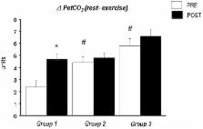
          A total of 123 patients (61.0±0.7 years) with coronary artery disease were divided according to aerobic fitness status into 3 groups: group 1 (n = 34, peak VO 2<17.5 ml/kg/min), group 2 (n = 67, peak VO 2>17.5 and <24.5 ml/kg/min) and group 3 (n = 22, peak VO 2>24.5 ml/kg/min). All patients performed a cardiorespiratory exercise test on a treadmill. Ventilatory efficiency was determined by the lowest VE/VCO 2 ratio observed. The exercise training program comprised moderate-intensity aerobic exercise performed 3 times per week for 3 months. Clinicaltrials.gov: NCT02106533

          RESULTS:

          Before intervention, group 1 exhibited both lower peak VO 2 and lower ventilatory efficiency compared with the other 2 groups ( p<0.05). After the exercise training program, group 1 exhibited greater improvements in aerobic fitness and ventilatory efficiency compared with the 2 other groups (group 1: ▵ = -2.5±0.5 units; group 2: ▵ = -0.8±0.3 units; and group 3: ▵ = -1.4±0.6 units, respectively; p<0.05).

          CONCLUSIONS:

          Coronary artery disease patients with lower aerobic fitness status exhibited lower ventilatory efficiency during a graded exercise test. In addition, after 3 months of aerobic exercise training, only the patients with initially lower levels of aerobic fitness exhibited greater improvements in ventilatory efficiency.

          Related collections

          Most cited references68

          • Record: found
          • Abstract: found
          • Article: not found

          Exercise pathophysiology in patients with primary pulmonary hypertension.

          Patients with primary pulmonary hypertension (PPH) have a pulmonary vasculopathy that leads to exercise intolerance due to dyspnea and fatigue. To better understand the basis of the exercise limitation in patients with PPH, cardiopulmonary exercise testing (CPET) with gas exchange measurements, New York Heart Association (NYHA) symptom class, and resting pulmonary hemodynamics were studied. We retrospectively evaluated 53 PPH patients who had right heart catheterization and cycle ergometer CPET studies to maximum tolerance as part of their clinical workups. No adverse events occurred during CPET. Reductions in peak O(2) uptake (VO(2)), anaerobic threshold, peak O(2) pulse, rate of increase in VO(2), and ventilatory efficiency were consistently found. NYHA class correlated well with the above parameters of aerobic function and ventilatory efficiency but less well with resting pulmonary hemodynamics. Patients with PPH can safely undergo noninvasive cycle ergometer CPET to their maximal tolerance. The CPET abnormalities were consistent and characteristic and correlated well with NYHA class.
            Bookmark
            • Record: found
            • Abstract: found
            • Article: not found

            Ventilatory efficiency during exercise in healthy subjects.

            When evaluating dyspnea in patients with heart or lung disease it is useful to measure the quantity of ventilation needed to eliminate metabolically produced CO2 (i.e., the ventilatory efficiency). Mathematically, the relationship between ventilation (VE) and CO2 output is determined by the arterial CO2 pressure and the physiologic dead space-tidal volume ratio. We decided to determine how age, sex, size, fitness, and the type of ergometer influenced ventilatory efficiency in normal subjects. Three methods were compared for expressing this relationship: (1) the VE versus CO2 output slope below the ventilatory compensation point, commonly used by cardiologists for estimating the severity of heart failure; (2) the VE/CO2 output ratio at the anaerobic threshold, commonly used by pulmonologists; and (3) the lowest VE/CO2 output ratio during exercise, the latter parameter not previously reported. We studied 474 healthy adults, between 17 and 78 years of age during incremental cycle and treadmill cardiopulmonary exercise tests at three test sites, correcting the total VE for the equipment dead space. The lowest VE/CO2 output ratio was insignificantly different from the ratio at the anaerobic threshold, less variable than that for the slope relationship, and unaffected by the site, ergometer, and gas exchange measurement systems. The regression equation for the lowest VE/CO2 output ratio was 27.94 + 0.108 x age + (0.97 = F, 0.0 = M) - 0.0376 x height, where age is in years and height is in centimeters. We conclude that the lowest VE/CO2 output ratio is the preferred noninvasive method to estimate ventilatory inefficiency.
              Bookmark
              • Record: found
              • Abstract: found
              • Article: not found

              Controlled trial of physical training in chronic heart failure. Exercise performance, hemodynamics, ventilation, and autonomic function.

              Many secondary abnormalities in chronic heart failure (CHF) may reflect physical deconditioning. There has been no prospective, controlled study of the effects of physical training on hemodynamics and autonomic function in CHF. In a controlled crossover trial of 8 weeks of exercise training, 17 men with stable moderate to severe CHF (age, 61.8 +/- 1.5 years; left ventricular ejection fraction, 19.6 +/- 2.3%), increased exercise tolerance (13.9 +/- 1.0 to 16.5 +/- 1.0 minutes, p less than 0.001), and peak oxygen uptake (13.2 +/- 0.9 to 15.6 +/- 1.0 ml/kg/min, p less than 0.01) significantly compared with controls. Training increased cardiac output at submaximal (5.9-6.7 l/min, p less than 0.05) and peak exercise (6.3-7.1 l/min, p less than 0.05), with a significant reduction in systemic vascular resistance. Training reduced minute ventilation and the slope relating minute ventilation to carbon dioxide production (-10.5%, p less than 0.05). Sympathovagal balance was altered by physical training when assessed by three methods: 1) RR variability (+19.2%, p less than 0.05); 2) autoregressive power spectral analysis of the resting ECG divided into low-frequency (-21.2%, p less than 0.01) and high-frequency (+51.3%, p less than 0.05) components; and 3) whole-body radiolabeled norepinephrine spillover (-16%, p less than 0.05). These measurements all showed a significant shift away from sympathetic toward enhanced vagal activity after training. Carefully selected patients with moderate to severe CHF can achieve significant, worthwhile improvements with exercise training. Physical deconditioning may be partly responsible for some of the associated abnormalities and exercise limitation of CHF, including abnormalities in autonomic balance.
                Bookmark

                Author and article information

                Journal
                Clinics (Sao Paulo)
                Clinics (Sao Paulo)
                Clinics
                Hospital das Clínicas da Faculdade de Medicina da Universidade de São Paulo
                1807-5932
                1980-5322
                January 2015
                : 70
                : 1
                : 46-51
                Affiliations
                [I ]Grupo Total Care, Amil, Cardiovascular Rehabilitation, São Paulo/SP, Brazil.
                [II ]Universidade Santa Cecília, Physical Education Department, Santos/SP, Brazil.
                Author notes

                Prado DM, Rocco EA and Silva AG conceived and designed the study. Prado DM, Rocco EA, Silva PF, Lazzari JM, Levefre G, Thies SB, Yamada CS, Puig RS and Furlan V acquired the data. Prado DM, Rocco EA and Silva AG analyzed and interpreted the data. All of the authors were involved in manuscript drafting or critically review for important intellectual content and all of the authors approved the manuscript final version for publication. Prado DM had full access to all of the data in the study and takes responsibility for the integrity of the data and the accuracy of the data analysis.

                *corresponding author
                Article
                cln_70p46
                10.6061/clinics/2015(01)09
                4311117
                25672429
                c86d85a3-e68c-4bdf-b187-5cadb7b3a25d
                Copyright © 2015 Hospital das Clínicas da FMUSP

                This is an Open Access article distributed under the terms of the Creative Commons Attribution Non-Commercial License ( http://creativecommons.org/licenses/by-nc/3.0/) which permits unrestricted non-commercial use, distribution, and reproduction in any medium, provided the original work is properly cited.

                History
                : 18 August 2014
                : 10 September 2014
                : 12 November 2014
                Page count
                Pages: 6
                Categories
                Clinical Science

                Medicine
                coronary disease,exercise,oxygen consumption,ventilation
                Medicine
                coronary disease, exercise, oxygen consumption, ventilation

                Comments

                Comment on this article