59
views
0
recommends
+1 Recommend
0 collections
    0
    shares
      • Record: found
      • Abstract: found
      • Article: found
      Is Open Access

      Structure and function of mitochondrial membrane protein complexes

      review-article
      BMC Biology
      BioMed Central

      Read this article at

      Bookmark
          There is no author summary for this article yet. Authors can add summaries to their articles on ScienceOpen to make them more accessible to a non-specialist audience.

          Abstract

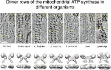
          Biological energy conversion in mitochondria is carried out by the membrane protein complexes of the respiratory chain and the mitochondrial ATP synthase in the inner membrane cristae. Recent advances in electron cryomicroscopy have made possible new insights into the structural and functional arrangement of these complexes in the membrane, and how they change with age. This review places these advances in the context of what is already known, and discusses the fundamental questions that remain open but can now be approached.

          Electronic supplementary material

          The online version of this article (doi:10.1186/s12915-015-0201-x) contains supplementary material, which is available to authorized users.

          Related collections

          Most cited references49

          • Record: found
          • Abstract: found
          • Article: not found

          Mitochondria as sensors and regulators of calcium signalling.

          During the past two decades calcium (Ca(2+)) accumulation in energized mitochondria has emerged as a biological process of utmost physiological relevance. Mitochondrial Ca(2+) uptake was shown to control intracellular Ca(2+) signalling, cell metabolism, cell survival and other cell-type specific functions by buffering cytosolic Ca(2+) levels and regulating mitochondrial effectors. Recently, the identity of mitochondrial Ca(2+) transporters has been revealed, opening new perspectives for investigation and molecular intervention.
            Bookmark
            • Record: found
            • Abstract: found
            • Article: not found

            Structure of the TRPV1 ion channel determined by electron cryo-microscopy

            Transient receptor potential (TRP) channels are sensors for a wide range of cellular and environmental signals, but elucidating how these channels respond to physical and chemical stimuli has been hampered by a lack of detailed structural information. Here, we exploit advances in electron cryo-microscopy to determine the structure of a mammalian TRP channel, TRPV1, at 3.4Å resolution, breaking the side-chain resolution barrier for membrane proteins without crystallization. Like voltage-gated channels, TRPV1 exhibits four-fold symmetry around a central ion pathway formed by transmembrane helices S5–S6 and the intervening pore loop, which is flanked by S1–S4 voltage sensor-like domains. TRPV1 has a wide extracellular ‘mouth’ with short selectivity filter. The conserved ‘TRP domain’ interacts with the S4–S5 linker, consistent with its contribution to allosteric modulation. Subunit organization is facilitated by interactions among cytoplasmic domains, including N-terminal ankyrin repeats. These observations provide a structural blueprint for understanding unique aspects of TRP channel function.
              Bookmark
              • Record: found
              • Abstract: found
              • Article: not found

              Cytochrome c and dATP-dependent formation of Apaf-1/caspase-9 complex initiates an apoptotic protease cascade.

              We report here the purification of the third protein factor, Apaf-3, that participates in caspase-3 activation in vitro. Apaf-3 was identified as a member of the caspase family, caspase-9. Caspase-9 and Apaf-1 bind to each other via their respective NH2-terminal CED-3 homologous domains in the presence of cytochrome c and dATP, an event that leads to caspase-9 activation. Activated caspase-9 in turn cleaves and activates caspase-3. Depletion of caspase-9 from S-100 extracts diminished caspase-3 activation. Mutation of the active site of caspase-9 attenuated the activation of caspase-3 and cellular apoptotic response in vivo, indicating that caspase-9 is the most upstream member of the apoptotic protease cascade that is triggered by cytochrome c and dATP.
                Bookmark

                Author and article information

                Contributors
                werner.kuehlbrandt@biophys.mpg.de
                Journal
                BMC Biol
                BMC Biol
                BMC Biology
                BioMed Central (London )
                1741-7007
                29 October 2015
                29 October 2015
                2015
                : 13
                : 89
                Affiliations
                Department of Structural Biology, Max Planck Institute of Biophysics, Max von Laue Str. 3, D-60438 Frankfurt am Main, Germany
                Article
                201
                10.1186/s12915-015-0201-x
                4625866
                26515107
                c9ed6a51-4142-4c88-9c17-96cf03849828
                © Kühlbrandt. 2015

                Open AccessThis article is distributed under the terms of the Creative Commons Attribution 4.0 International License ( http://creativecommons.org/licenses/by/4.0/), which permits unrestricted use, distribution, and reproduction in any medium, provided you give appropriate credit to the original author(s) and the source, provide a link to the Creative Commons license, and indicate if changes were made. The Creative Commons Public Domain Dedication waiver ( http://creativecommons.org/publicdomain/zero/1.0/) applies to the data made available in this article, unless otherwise stated.

                History
                Categories
                Review
                Custom metadata
                © The Author(s) 2015

                Life sciences
                Life sciences

                Comments

                Comment on this article

                scite_

                Similar content162

                Cited by182

                Most referenced authors1,319