21
views
0
recommends
+1 Recommend
0 collections
    0
    shares
      • Record: found
      • Abstract: found
      • Article: found
      Is Open Access

      A Case of Multiple Cardiovascular and Tracheal Anomalies Presented with Wolff-Parkinson-White Syndrome in a Middle-aged Adult

      case-report

      Read this article at

      Bookmark
          There is no author summary for this article yet. Authors can add summaries to their articles on ScienceOpen to make them more accessible to a non-specialist audience.

          Abstract

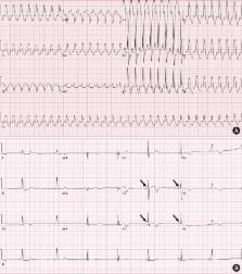
          Congenital cardiovascular anomalies, such as dextrocardia, persistent left superior vena cava (SVC), and pulmonary artery (PA) sling, are rare disorders. These congenital anomalies can occur alone, or coincide with other congenital malformations. In the majority of cases, congenital anomalies are detected early in life by certain signs and symptoms. A 56-year-old man with no previous medical history was admitted due to recurrent wide QRS complex tachycardia with hemodynamic collapse. A chest radiograph showed dextrocardia. After synchronized cardioversion, an electrocardiogram revealed Wolff-Parkinson-White (WPW) syndrome. Persistent left SVC, PA sling, and right tracheal bronchus were also detected by a chest computed tomography (CT) scan. He was diagnosed with paroxysmal supraventricular tachycardia (PSVT) associated with WPW syndrome, and underwent radiofrequency ablation. We reported the first case of situs solitus dextrocardia coexisting with persistent left SVC, PA sling and right tracheal bronchus presented with WPW and PSVT in a middle-aged adult. In patients with a cardiovascular anomaly, clinicians should consider thorough evaluation of possibly combined cardiovascular and airway malformations and cardiac dysrhythmia.

          Graphical Abstract

          Related collections

          Most cited references12

          • Record: found
          • Abstract: found
          • Article: not found

          A population-based study of cardiac malformations and outcomes associated with dextrocardia.

          The incidence of dextrocardia and its associated cardiac and noncardiac malformations is not known. There is inadequate information about outcomes to counsel parents about prognosis. A retrospective review of all diagnoses of dextrocardia due to embryologic development at a tertiary care hospital from 1985 to 2001 was performed. Eighty-one cases were identified (48 antenatally). The incidence of dextrocardia was estimated to be 1 in 12,019 pregnancies. Twenty-seven cases were situs solitus, 30 situs inversus, and 24 situs ambiguous or isomerism. Cardiac malformations were found in 26 of 27 cases of situs solitus, 7 of 30 cases of situs inversus, and 24 of 24 cases of isomerism. Noncardiac malformations were identified in 10 of 27 cases of situs solitus, 6 of 30 cases of situs inversus, and 14 of 24 cases of isomerism. Twelve pregnancies were terminated, 3 fetuses were stillborn, and 2 women chose compassionate care. All terminated fetuses were diagnosed with dextrocardia before termination, and all had >1 cardiac anomaly; 7 also had noncardiac anomalies. There were 43 subjects in the intention-to-treat group (20 situs solitus, 10 solitus inversus, 13 isomerism). Thirty-two had >or=1 cardiac operation, and 21 had >or=3. Thirty-nine subjects were alive at most recent follow-up. In conclusion, the incidence of dextrocardia was 1 in 12,019 pregnancies. In conclusion, in our cohort, the numbers of cases of situs solitus, situs inversus, and isomerism were similar. Cardiac and noncardiac malformations were most common in the isomerism group. Cardiac malformations were often complex in the situs solitus and isomerism groups. Ninety-one percent of those in the intention-to-treat cohort were alive at follow-up.
            Bookmark
            • Record: found
            • Abstract: found
            • Article: not found

            Approach to dextrocardia in adults: review.

            The educational objectives of this article are to describe an approach to analyzing imaging studies in adults with dextrocardia and to present the appearances of the most common underlying disorders. Topics reviewed include the morphology of the cardiac chambers, the concept of situs, and the relevant embryologic principles. The disorders discussed include situs inversus totalis (mirror-image dextrocardia), dextroversion, congenitally corrected transposition of the great arteries, and polysplenia syndrome.
              Bookmark
              • Record: found
              • Abstract: found
              • Article: not found

              Persistent left superior vena cava draining into the coronary sinus: report of 10 cases and literature review.

              Persistent left superior vena cava (PLSVC) is the most common thoracic venous anomaly. This study examines the epidemiologic, clinical, and morphologic characteristics of a cohort of patients with PLSVC draining into the coronary sinus. We examined the clinical and morphologic characteristics of patients with PLSVC draining into the coronary sinus diagnosed at a single referral hospital for a defined population in northwestern Spain. We designed a prospective study of the case records of all patients diagnosed with PLSVC draining into the coronary sinus at the echocardiography laboratory of the Hospital Xeral-Calde from January 2001 through December 2002. Patients were included if they had a PLSVC diagnosed by transthoracic echocardiogram (TTE) using an echo-contrast enhancement and confirmed by a magnetic resonance (MR) imaging. Ten patients (6 women) fulfilled the inclusion criteria described above. All patients were adults and had associated heart disease, including a congenital heart disease in three cases. Magnetic resonance imaging examination confirmed the presence of PLSVC and the site of drainage into the coronary sinus. Absence of the right superior vena cava was observed only in three patients, in whom the main coronary sinus size was significantly increased. Absence of the left brachiocephalic vein was diagnosed in five patients. This study describes 10 new cases of PLSVC and supports the necessity of considering PLSVC draining into the coronary sinus in the diagnosis of patients presenting with dilated coronary sinus diagnosed by TTE. It also underlines the important role of MR imaging in the evaluation of these abnormalities. An associated heart disease must always be excluded in these patients.
                Bookmark

                Author and article information

                Journal
                J Korean Med Sci
                J. Korean Med. Sci
                JKMS
                Journal of Korean Medical Science
                The Korean Academy of Medical Sciences
                1011-8934
                1598-6357
                December 2017
                28 September 2017
                : 32
                : 12
                : 2069-2072
                Affiliations
                [1 ]Department of Internal Medicine, Hallym University Kangdong Sacred Heart Hospital, Seoul, Korea.
                [2 ]Department of Internal Medicine, Jeju National University School of Medicine, Jeju National University Hospital, Jeju, Korea.
                [3 ]Department of Radiology, Jeju National University School of Medicine, Jeju National University Hospital, Jeju, Korea.
                Author notes
                Address for Correspondence: Changhwan Kim, MD. Department of Internal Medicine, Jeju National University School of Medicine, Jeju National University Hospital, 15 Aran 13-gil, Jeju 63241, Republic of Korea. masque70@ 123456dreamwiz.com
                Author information
                https://orcid.org/0000-0003-2155-6430
                https://orcid.org/0000-0001-7840-4767
                https://orcid.org/0000-0003-2315-8129
                https://orcid.org/0000-0001-6554-1316
                https://orcid.org/0000-0001-9130-0997
                https://orcid.org/0000-0002-8466-7501
                https://orcid.org/0000-0001-5277-2254
                https://orcid.org/0000-0003-3318-2528
                Article
                10.3346/jkms.2017.32.12.2069
                5680510
                29115093
                cc51018c-89c0-49aa-8b43-fe22b8c24900
                © 2017 The Korean Academy of Medical Sciences.

                This is an Open Access article distributed under the terms of the Creative Commons Attribution Non-Commercial License ( https://creativecommons.org/licenses/by-nc/4.0/) which permits unrestricted non-commercial use, distribution, and reproduction in any medium, provided the original work is properly cited.

                History
                : 27 March 2016
                : 13 August 2016
                Categories
                Case Report
                Cardiovascular Disorders

                Medicine
                dextrocardia,superior vena cava,pulmonary artery,bronchus,wolff-parkinson-white syndrome
                Medicine
                dextrocardia, superior vena cava, pulmonary artery, bronchus, wolff-parkinson-white syndrome

                Comments

                Comment on this article