16
views
0
recommends
+1 Recommend
0 collections
    0
    shares
      • Record: found
      • Abstract: found
      • Article: found
      Is Open Access

      Reduced TET2 function leads to T-cell lymphoma with follicular helper T-cell-like features in mice

      research-article

      Read this article at
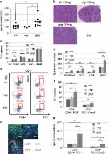
      Bookmark
          There is no author summary for this article yet. Authors can add summaries to their articles on ScienceOpen to make them more accessible to a non-specialist audience.

          Abstract

          TET2 (Ten Eleven Translocation 2) is a dioxygenase that converts methylcytosine (mC) to hydroxymethylcytosine (hmC). TET2 loss-of-function mutations are highly frequent in subtypes of T-cell lymphoma that harbor follicular helper T (Tfh)-cell-like features, such as angioimmunoblastic T-cell lymphoma (30–83%) or peripheral T-cell lymphoma, not otherwise specified (10–49%), as well as myeloid malignancies. Here, we show that middle-aged Tet2 knockdown ( Tet2 gt/gt ) mice exhibit Tfh-like cell overproduction in the spleen compared with control mice. The Tet2 knockdown mice eventually develop T-cell lymphoma with Tfh-like features after a long latency (median 67 weeks). Transcriptome analysis revealed that these lymphoma cells had Tfh-like gene expression patterns when compared with splenic CD4-positive cells of wild-type mice. The lymphoma cells showed lower hmC densities around the transcription start site (TSS) and higher mC densities at the regions of the TSS, gene body and CpG islands. These epigenetic changes, seen in Tet2 insufficiency-triggered lymphoma, possibly contributed to predated outgrowth of Tfh-like cells and subsequent lymphomagenesis. The mouse model described here suggests that TET2 mutations play a major role in the development of T-cell lymphoma with Tfh-like features in humans.

          Related collections

          Most cited references26

          • Record: found
          • Abstract: found
          • Article: not found

          Efficacy of azacitidine compared with that of conventional care regimens in the treatment of higher-risk myelodysplastic syndromes: a randomised, open-label, phase III study.

          Drug treatments for patients with high-risk myelodysplastic syndromes provide no survival advantage. In this trial, we aimed to assess the effect of azacitidine on overall survival compared with the three commonest conventional care regimens. In a phase III, international, multicentre, controlled, parallel-group, open-label trial, patients with higher-risk myelodysplastic syndromes were randomly assigned one-to-one to receive azacitidine (75 mg/m(2) per day for 7 days every 28 days) or conventional care (best supportive care, low-dose cytarabine, or intensive chemotherapy as selected by investigators before randomisation). Patients were stratified by French-American-British and international prognostic scoring system classifications; randomisation was done with a block size of four. The primary endpoint was overall survival. Efficacy analyses were by intention to treat for all patients assigned to receive treatment. This study is registered with ClinicalTrials.gov, number NCT00071799. Between Feb 13, 2004, and Aug 7, 2006, 358 patients were randomly assigned to receive azacitidine (n=179) or conventional care regimens (n=179). Four patients in the azacitidine and 14 in the conventional care groups received no study drugs but were included in the intention-to-treat efficacy analysis. After a median follow-up of 21.1 months (IQR 15.1-26.9), median overall survival was 24.5 months (9.9-not reached) for the azacitidine group versus 15.0 months (5.6-24.1) for the conventional care group (hazard ratio 0.58; 95% CI 0.43-0.77; stratified log-rank p=0.0001). At last follow-up, 82 patients in the azacitidine group had died compared with 113 in the conventional care group. At 2 years, on the basis of Kaplan-Meier estimates, 50.8% (95% CI 42.1-58.8) of patients in the azacitidine group were alive compared with 26.2% (18.7-34.3) in the conventional care group (p<0.0001). Peripheral cytopenias were the most common grade 3-4 adverse events for all treatments. Treatment with azacitidine increases overall survival in patients with higher-risk myelodysplastic syndromes relative to conventional care.
            Bookmark
            • Record: found
            • Abstract: found
            • Article: not found

            Genome-wide mapping of 5-hydroxymethylcytosine in embryonic stem cells.

            5-hydroxymethylcytosine (5hmC) is a modified base present at low levels in diverse cell types in mammals. 5hmC is generated by the TET family of Fe(II) and 2-oxoglutarate-dependent enzymes through oxidation of 5-methylcytosine (5mC). 5hmC and TET proteins have been implicated in stem cell biology and cancer, but information on the genome-wide distribution of 5hmC is limited. Here we describe two novel and specific approaches to profile the genomic localization of 5hmC. The first approach, termed GLIB (glucosylation, periodate oxidation, biotinylation) uses a combination of enzymatic and chemical steps to isolate DNA fragments containing as few as a single 5hmC. The second approach involves conversion of 5hmC to cytosine 5-methylenesulphonate (CMS) by treatment of genomic DNA with sodium bisulphite, followed by immunoprecipitation of CMS-containing DNA with a specific antiserum to CMS. High-throughput sequencing of 5hmC-containing DNA from mouse embryonic stem (ES) cells showed strong enrichment within exons and near transcriptional start sites. 5hmC was especially enriched at the start sites of genes whose promoters bear dual histone 3 lysine 27 trimethylation (H3K27me3) and histone 3 lysine 4 trimethylation (H3K4me3) marks. Our results indicate that 5hmC has a probable role in transcriptional regulation, and suggest a model in which 5hmC contributes to the 'poised' chromatin signature found at developmentally-regulated genes in ES cells.
              Bookmark
              • Record: found
              • Abstract: found
              • Article: not found

              Uncovering the role of 5-hydroxymethylcytosine in the epigenome.

              Just over 2 years ago, TET1 was found to catalyse the oxidation of 5-methylcytosine, a well-known epigenetic mark, into 5-hydroxymethylcytosine in mammalian DNA. The exciting prospect of a novel epigenetic modification that may dynamically regulate DNA methylation has led to the rapid accumulation of publications from a wide array of fields, from biochemistry to stem cell biology. Although we have only started to scratch the surface, interesting clues on the role of 5-hydroxymethylcytosine are quickly emerging.
                Bookmark

                Author and article information

                Journal
                Blood Cancer J
                Blood Cancer J
                Blood Cancer Journal
                Nature Publishing Group
                2044-5385
                December 2014
                12 December 2014
                1 December 2014
                : 4
                : 12
                : e264
                Affiliations
                [1 ]Department of Hematology, University of Tsukuba Hospital, Tsukuba, Ibaraki, Japan
                [2 ]Graduate School of Comprehensive Human Science, University of Tsukuba , Tsukuba, Ibaraki, Japan
                [3 ]Faculty of Medicine, Department of Hematology, University of Tsukuba , Tsukuba, Ibaraki, Japan
                [4 ]Genome Science Division, Research Center for Advanced Science and Technology, The University of Tokyo , Meguro-ku, Tokyo, Japan
                [5 ]Department of Pathology and Tumor Biology, Graduate School of Medicine, Kyoto University , Kyoto, Japan
                [6 ]Life Science center, Tsukuba Advanced Research Alliance, University of Tsukuba , Tsukuba, Ibaraki, Japan
                [7 ]Department of Molecular Biological Oncology, Faculty of Medicine, University of Tsukuba , Tsukuba, Ibaraki, Japan
                [8 ]Department of Clinical Laboratory, University of Tsukuba Hospital , Tsukuba, Ibaraki, Japan
                [9 ]Department of Pathology, Tokai University School of Medicine , Isehara, Kanagawa, Japan
                Author notes
                [* ]Department of Hematology, Faculty of Medicine, University of Tsukuba , 1-1-1 Tennodai, Tsukuba 305-8575, Ibaraki, Japan. E-mail: schiba-t@ 123456md.tsukuba.ac.jp
                Article
                bcj201483
                10.1038/bcj.2014.83
                4315889
                25501021
                cc828fbd-187f-4f4b-b9b7-40a2e51bad09
                Copyright © 2014 Macmillan Publishers Limited

                This work is licensed under a Creative Commons Attribution-NonCommercial-NoDerivs 4.0 International License. The images or other third party material in this article are included in the article's Creative Commons license, unless indicated otherwise in the credit line; if the material is not included under the Creative Commons license, users will need to obtain permission from the license holder to reproduce the material. To view a copy of this license, visit http://creativecommons.org/licenses/by-nc-nd/4.0/

                History
                : 30 September 2014
                : 02 October 2014
                Categories
                Original Article

                Oncology & Radiotherapy
                Oncology & Radiotherapy

                Comments

                Comment on this article