4
views
0
recommends
+1 Recommend
0 collections
    0
    shares
      • Record: found
      • Abstract: found
      • Article: found
      Is Open Access

      Prostaglandin E 2 stimulates the epithelial sodium channel (ENaC) in cultured mouse cortical collecting duct cells in an autocrine manner

      research-article

      Read this article at

      Bookmark
          There is no author summary for this article yet. Authors can add summaries to their articles on ScienceOpen to make them more accessible to a non-specialist audience.

          Abstract

          In murine cortical collecting duct cells, prostaglandin E 2 (PGE 2) stimulates transepithelial sodium transport mediated by the epithelial sodium channel (ENaC). PGE 2 is synthesized and secreted by the cells and acts on basolateral prostaglandin E receptor type 4 (EP 4).

          Abstract

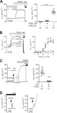
          Prostaglandin E 2 (PGE 2) is the most abundant prostanoid in the kidney, affecting a wide range of renal functions. Conflicting data have been reported regarding the effects of PGE 2 on tubular water and ion transport. The amiloride-sensitive epithelial sodium channel (ENaC) is rate limiting for transepithelial sodium transport in the aldosterone-sensitive distal nephron. The aim of the present study was to explore a potential role of PGE 2 in regulating ENaC in cortical collecting duct (CCD) cells. Short-circuit current (I SC) measurements were performed using the murine mCCD cl1 cell line known to express characteristic properties of CCD principal cells and to be responsive to physiological concentrations of aldosterone and vasopressin. PGE 2 stimulated amiloride-sensitive I SC via basolateral prostaglandin E receptors type 4 (EP 4) with an EC 50 of ∼7.1 nM. The rapid stimulatory effect of PGE 2 on I SC resembled that of vasopressin. A maximum response was reached within minutes, coinciding with an increased abundance of β-ENaC at the apical plasma membrane and elevated cytosolic cAMP levels. The effects of PGE 2 and vasopressin were nonadditive, indicating similar signaling cascades. Exposing mCCD cl1 cells to aldosterone caused a much slower (∼2 h) increase of the amiloride-sensitive I SC. Interestingly, the rapid effect of PGE 2 was preserved even after aldosterone stimulation. Furthermore, application of arachidonic acid also increased the amiloride-sensitive I SC involving basolateral EP 4 receptors. Exposure to arachidonic acid resulted in elevated PGE 2 in the basolateral medium in a cyclooxygenase 1 (COX-1)–dependent manner. These data suggest that in the cortical collecting duct, locally produced and secreted PGE 2 can stimulate ENaC-mediated transepithelial sodium transport.

          Related collections

          Most cited references91

          • Record: found
          • Abstract: found
          • Article: not found

          Prostanoid receptors: structures, properties, and functions.

          Prostanoids are the cyclooxygenase metabolites of arachidonic acid and include prostaglandin (PG) D(2), PGE(2), PGF(2alpha), PGI(2), and thromboxne A(2). They are synthesized and released upon cell stimulation and act on cells in the vicinity of their synthesis to exert their actions. Receptors mediating the actions of prostanoids were recently identified and cloned. They are G protein-coupled receptors with seven transmembrane domains. There are eight types and subtypes of prostanoid receptors that are encoded by different genes but as a whole constitute a subfamily in the superfamily of the rhodopsin-type receptors. Each of the receptors was expressed in cultured cells, and its ligand-binding properties and signal transduction pathways were characterized. Moreover, domains and amino acid residues conferring the specificities of ligand binding and signal transduction are being clarified. Information also is accumulating as to the distribution of these receptors in the body. It is also becoming clear for some types of receptors how expression of their genes is regulated. Furthermore, the gene for each of the eight types of prostanoid receptor has been disrupted, and mice deficient in each type of receptor are being examined to identify and assess the roles played by each receptor under various physiological and pathophysiological conditions. In this article, we summarize these findings and attempt to give an overview of the current status of research on the prostanoid receptors.
            Bookmark
            • Record: found
            • Abstract: found
            • Article: not found

            Epithelial sodium channels: function, structure, and regulation.

            The apical (outward-facing) membranes of high-resistance epithelia contain Na+ channels, traditionally identified by their sensitivity to block by the K(+)-sparing diuretic amiloride. Such channels have been characterized in amphibian skin and urinary bladder, renal collecting duct, distal colon, sweat and salivary glands, lung, and taste buds. They mediate the first step of active Na+ reabsorption and play a major role in the maintenance of electrolyte and water homeostasis in all vertebrates. In the past, these channels were classified according to their biophysical and pharmacological properties. The recent cloning of the three homologous channel subunits denoted alpha-, beta-, and gamma-epithelial Na+ channels (ENaC) has provided a molecular definition of at least one class of amiloride-blockable channels. Subsequent studies have established that ENaC is a major Na(+)-conducting pathway in both absorbing and secretory epithelia and is related to one type of channel involved in mechanosensation. This review summarizes the biophysical characteristics, molecular properties, and regulatory mechanisms of epithelial amiloride-blockable Na+ channels. Special emphasis is given to recent studies utilizing cloned ENaC subunits and purified amiloride-binding proteins.
              Bookmark
              • Record: found
              • Abstract: found
              • Article: not found

              Single-Cell Profiling Reveals Sex, Lineage, and Regional Diversity in the Mouse Kidney

              Chronic kidney disease affects 10% of the population with notable differences in ethnic and sex-related susceptibility to kidney injury and disease. Kidney dysfunction leads to significant morbidity and mortality, and chronic disease in other organ systems. A mouse organ-centered understanding underlies rapid progress in human disease modeling and cellular approaches to repair damaged systems. To enhance an understanding of the mammalian kidney, we combined anatomy-guided single cell RNA sequencing of the adult male and female mouse kidney with in situ expression studies and cell lineage tracing. These studies reveal cell diversity and marked sex differences, distinct organization and cell composition of nephrons dependent on the time of nephron specification, and lineage convergence, in which contiguous functionally-related cell types are specified from nephron and collecting system progenitor populations. A searchable database, Kidney Cell Explorer ( https://cello.shinyapps.io/kidneycellexplorer/ ), enables gene-cell relationships to be viewed in the anatomical framework of the kidney. Ransick et al. combined in depth single cell profiling of male and female adult kidneys with cluster registration to kidney structures to generate an anatomical atlas of the mammalian nephron and collecting system. Kidney Cell Explorer (( https://cello.shinyapps.io/kidneycellexplorer/ ) enables gene-cell relationships to be viewed in the anatomical framework of the mammalian kidney.
                Bookmark

                Author and article information

                Journal
                J Gen Physiol
                J. Gen. Physiol
                jgp
                The Journal of General Physiology
                Rockefeller University Press
                0022-1295
                1540-7748
                03 August 2020
                22 May 2020
                : 152
                : 8
                : e201912525
                Affiliations
                [1 ]Institute of Cellular and Molecular Physiology, Friedrich-Alexander University Erlangen-Nürnberg, Erlangen, Germany
                [2 ]Institute of Experimental and Clinical Pharmacology and Toxicology, Friedrich-Alexander University Erlangen-Nürnberg, Erlangen, Germany
                Author notes
                Correspondence to Christoph Korbmacher: christoph.korbmacher@ 123456fau.de
                [*]

                M.K. Mansley and C. Niklas contributed equally to this paper.

                Author information
                https://orcid.org/0000-0002-8082-0273
                https://orcid.org/0000-0002-8887-0996
                https://orcid.org/0000-0003-2127-4185
                https://orcid.org/0000-0002-1504-9582
                Article
                jgp.201912525
                10.1085/jgp.201912525
                7398144
                32442241
                cd2fcbfc-3d89-461f-837a-5fa6b96edbec
                © 2020 Mansley et al.

                This article is distributed under the terms of an Attribution–Noncommercial–Share Alike–No Mirror Sites license for the first six months after the publication date (see http://www.rupress.org/terms/). After six months it is available under a Creative Commons License (Attribution–Noncommercial–Share Alike 4.0 International license, as described at https://creativecommons.org/licenses/by-nc-sa/4.0/).

                History
                : 02 November 2019
                : 30 March 2020
                : 21 April 2020
                Page count
                Pages: 15
                Funding
                Funded by: Deutsche Forschungsgemeinschaft, DOI http://dx.doi.org/10.13039/501100001659;
                Award ID: SFB 1350
                Funded by: Alexander von Humboldt Foundation, DOI http://dx.doi.org/10.13039/100005156;
                Award ID: 3.3-GRO/1143730 STP
                Funded by: Interdisziplinäres Zentrum für Klinische Forschung, DOI http://dx.doi.org/10.13039/501100009379;
                Award ID: IZKF, TP-A33
                Funded by: Bayerische Forschungsstiftung, DOI http://dx.doi.org/10.13039/501100002745;
                Award ID: PDOK-74-10
                Categories
                Article
                Membrane Transport

                Anatomy & Physiology
                Anatomy & Physiology

                Comments

                Comment on this article