2
views
0
recommends
+1 Recommend
0 collections
    0
    shares
      • Record: found
      • Abstract: found
      • Article: found
      Is Open Access

      High Serum Uric Acid is Highly Associated with a Reduced Left Ventricular Ejection Fraction Rather than Increased Plasma B-type Natriuretic Peptide in Patients with Cardiovascular Diseases

      research-article

      Read this article at

      Bookmark
          There is no author summary for this article yet. Authors can add summaries to their articles on ScienceOpen to make them more accessible to a non-specialist audience.

          Abstract

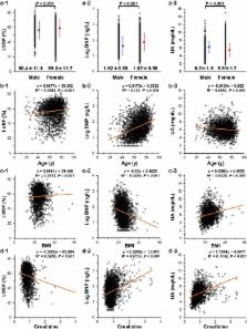
          High serum uric acid (UA) has been reported to be associated with left ventricular (LV) dysfunction; however, the relationship between UA and plasma B-type natriuretic peptide (BNP), a sensitive biomarker of heart failure, is still unclear. This study investigated their relationship to provide an accurate assessment of high UA. The study patients consisted of 3,077 subjects who underwent cardiac catheterization because of various cardiovascular disorders. Since the explanatory factors of multiple regression analysis were mostly confounding with each other, subgroup analysis was performed by quartering the study population using the respective risk factors and by covariance structure analysis. This analysis revealed that UA was almost always well associated with a reduced LV ejection fraction (LVEF), but generally not with BNP. UA was significantly associated with BNP in lean aged females, but not in obese adolescent males, although LVEF was significantly reduced in response to a high UA in both groups. A high UA is a direct risk factor for cardiac dysfunction from the perspective of BNP; however, augmentation of BNP in response to a high UA would likely be restricted among obese adolescent males. On the other hand, the observed LV systolic dysfunction, such as LVEF, reflects a high UA on an almost constant basis.

          Related collections

          Most cited references41

          • Record: found
          • Abstract: found
          • Article: not found

          A role for uric acid in the progression of renal disease.

          Hyperuricemia is associated with renal disease, but it is usually considered a marker of renal dysfunction rather than a risk factor for progression. Recent studies have reported that mild hyperuricemia in normal rats induced by the uricase inhibitor, oxonic acid (OA), results in hypertension, intrarenal vascular disease, and renal injury. This led to the hypothesis that uric acid may contribute to progressive renal disease. To examine the effect of hyperuricemia on renal disease progression, rats were fed 2% OA for 6 wk after 5/6 remnant kidney (RK) surgery with or without the xanthine oxidase inhibitor, allopurinol, or the uricosuric agent, benziodarone. Renal function and histologic studies were performed at 6 wk. Given observations that uric acid induces vascular disease, the effect of uric acid on vascular smooth muscle cells in culture was also examined. RK rats developed transient hyperuricemia (2.7 mg/dl at week 2), but then levels returned to baseline by week 6 (1.4 mg/dl). In contrast, RK+OA rats developed higher and more persistent hyperuricemia (6 wk, 3.2 mg/dl). Hyperuricemic rats demonstrated higher BP, greater proteinuria, and higher serum creatinine than RK rats. Hyperuricemic RK rats had more renal hypertrophy and greater glomerulosclerosis (24.2 +/- 2.5 versus 17.5 +/- 3.4%; P < 0.05) and interstitial fibrosis (1.89 +/- 0.45 versus 1.52 +/- 0.47; P < 0.05). Hyperuricemic rats developed vascular disease consisting of thickening of the preglomerular arteries with smooth muscle cell proliferation; these changes were significantly more severe than a historical RK group with similar BP. Allopurinol significantly reduced uric acid levels and blocked the renal functional and histologic changes. Benziodarone reduced uric acid levels less effectively and only partially improved BP and renal function, with minimal effect on the vascular changes. To better understand the mechanism for the vascular disease, the expression of COX-2 and renin were examined. Hyperuricemic rats showed increased renal renin and COX-2 expression, the latter especially in preglomerular arterial vessels. In in vitro studies, cultured vascular smooth muscle cells incubated with uric acid also generated COX-2 with time-dependent proliferation, which was prevented by either a COX-2 or TXA-2 receptor inhibitor. Hyperuricemia accelerates renal progression in the RK model via a mechanism linked to high systemic BP and COX-2-mediated, thromboxane-induced vascular disease. These studies provide direct evidence that uric acid may be a true mediator of renal disease and progression.
            Bookmark
            • Record: found
            • Abstract: found
            • Article: found
            Is Open Access

            Association Between Serum Uric Acid and Development of Type 2 Diabetes

            OBJECTIVE To systematically evaluate the association between serum uric acid (SUA) level and subsequent development of type 2 diabetes. RESEARCH DESIGN AND METHODS We searched Medline (31 March from 1966 to 2009) and Embase (31 March from 1980 to 2009) for observational cohort studies examining the association between SUA and the risk of type 2 diabetes by manual literature search. Relative risks (RRs) for each 1 mg/dl increase in SUA were pooled by using a random-effects model. The studies included were stratified into subgroups representing different study characteristics, and meta-regression analyses were performed to investigate the effect of these characteristics on the association between SUA level and type 2 diabetes risk. RESULTS The search yielded 11 cohort studies (42,834 participants) that reported 3,305 incident cases of type 2 diabetes during follow-up periods ranging from 2.0 to 13.5 years. The pooled RR of a 1 mg/dl increase in SUA was 1.17 (95% CI 1.09–1.25). Study results were consistently significant (i.e., >1) across characteristics of participants and study design. Publication bias was both visually and statistically suggested (P = 0.03 for Egger's test, 0.06). Adjustment for publication bias attenuated the pooled RR per mg/dl increase in SUA (RR 1.11 [95% CI 1.03–1.20]), but the association remained statistically significant (P = 0.009). CONCLUSIONS The current meta-analysis suggests that SUA level is positively associated with the development of type 2 diabetes regardless of various study characteristics. Further research should attempt to determine whether it is effective to utilize SUA level as a predictor of type 2 diabetes for its primary prevention.
              Bookmark
              • Record: found
              • Abstract: found
              • Article: not found

              Brain natriuretic peptide as a novel cardiac hormone in humans. Evidence for an exquisite dual natriuretic peptide system, atrial natriuretic peptide and brain natriuretic peptide.

              Using a specific radioimmunoassay for human brain natriuretic peptide (hBNP) with a monoclonal antibody, we have investigated its synthesis, secretion, and clearance in comparison with those of atrial natriuretic peptide (ANP) in normal subjects and patients with congestive heart failure (CHF). Mean BNP-like immunoreactivity (-LI) levels in normal atrium and ventricle were 250 and 18 pmol/g, respectively. The plasma BNP-LI level in normal subjects was 0.90 +/- 0.07 fmol/ml, which was 16% of the ANP-LI level. In contrast, the plasma BNP-LI level markedly increased in patients with CHF in proportion to its severity, and surpassed the ANP-LI level in severe cases. There was a significant step-up of the plasma BNP-LI level in the coronary sinus (CS) compared with that in the aortic root (Ao) and the difference between these BNP-LI levels, delta(CS-Ao)BNP, also increased with the severity of CHF. In addition, the step-up of the BNP-LI level in the anterior interventricular vein [delta(AIV-Ao)BNP] was comparable to delta(CS-Ao)BNP, indicating that BNP is secreted mainly from the ventricle. Predominant BNP synthesis in the ventricle was also confirmed by Northern blot analysis. Catheterization and pharmacokinetic studies revealed that hBNP is cleared from the circulation more slowly than alpha-hANP; this was in part attributed to lower (about 7%) binding affinity of hBNP to clearance receptors than that of alpha-hANP. A predominant molecular form of BNP-LI in the heart and plasma was a 3-kD form corresponding to hBNP. These results indicate that BNP is a novel cardiac hormone secreted predominantly from the ventricle, and that the synthesis, secretion and clearance of BNP differ from those of ANP, suggesting discrete physiological and pathophysiological roles of BNP in a dual natriuretic peptide system.
                Bookmark

                Author and article information

                Contributors
                cadmk@jikei.ac.jp
                Journal
                Sci Rep
                Sci Rep
                Scientific Reports
                Nature Publishing Group UK (London )
                2045-2322
                24 January 2019
                24 January 2019
                2019
                : 9
                : 682
                Affiliations
                ISNI 0000 0001 0661 2073, GRID grid.411898.d, Division of Cardiology, Department of Internal Medicine, , The Jikei University School of Medicine, ; Tokyo, Japan
                Author information
                http://orcid.org/0000-0002-0914-9376
                Article
                37053
                10.1038/s41598-018-37053-0
                6346056
                30679647
                cd3d5cff-5c13-42e4-a8b8-619f5e14d410
                © The Author(s) 2019

                Open Access This article is licensed under a Creative Commons Attribution 4.0 International License, which permits use, sharing, adaptation, distribution and reproduction in any medium or format, as long as you give appropriate credit to the original author(s) and the source, provide a link to the Creative Commons license, and indicate if changes were made. The images or other third party material in this article are included in the article’s Creative Commons license, unless indicated otherwise in a credit line to the material. If material is not included in the article’s Creative Commons license and your intended use is not permitted by statutory regulation or exceeds the permitted use, you will need to obtain permission directly from the copyright holder. To view a copy of this license, visit http://creativecommons.org/licenses/by/4.0/.

                History
                : 21 August 2018
                : 30 November 2018
                Categories
                Article
                Custom metadata
                © The Author(s) 2019

                Uncategorized
                Uncategorized

                Comments

                Comment on this article