57
views
0
recommends
+1 Recommend
0 collections
    0
    shares
      • Record: found
      • Abstract: found
      • Article: not found

      The Role of HDAC6 in Cancer

      review-article

      Read this article at

      ScienceOpenPublisherPMC
      Bookmark
          There is no author summary for this article yet. Authors can add summaries to their articles on ScienceOpen to make them more accessible to a non-specialist audience.

          Abstract

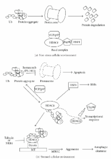
          Histone deacetylase 6 (HDAC6), a member of the HDAC family whose major substrate is α-tubulin, has become a target for drug development to treat cancer due to its major contribution in oncogenic cell transformation. Overexpression of HDAC6 correlates with tumorigenesis and improved survival; therefore, HDAC6 may be used as a marker for prognosis. Previous work demonstrated that in multiple myeloma cells, inhibition of HDAC6 results in apoptosis. Furthermore, HDAC6 is required for the activation of heat-shock factor 1 (HSF1), an activator of heat-shock protein encoding genes (HSPs) and CYLD, a cylindromatosis tumor suppressor gene. HDAC6 contributes to cancer metastasis since its upregulation increases cell motility in breast cancer MCF-7 cells and its interaction with cortactin regulates motility. HDAC6 also affects transcription and translation by regulating the heat-shock protein 90 (Hsp90) and stress granules (SGs), respectively. This review will discuss the role of HDAC6 in the pathogenesis and treatment of cancer.

          Related collections

          Most cited references49

          • Record: found
          • Abstract: found
          • Article: not found

          Histone deacetylases and cancer: causes and therapies.

          Together, histone acetyltransferases and histone deacetylases (HDACs) determine the acetylation status of histones. This acetylation affects the regulation of gene expression, and inhibitors of HDACs have been found to cause growth arrest, differentiation and/or apoptosis of many tumours cells by altering the transcription of a small number of genes. HDAC inhibitors are proving to be an exciting therapeutic approach to cancer, but how do they exert this effect?
            Bookmark
            • Record: found
            • Abstract: found
            • Article: not found

            Structures of a histone deacetylase homologue bound to the TSA and SAHA inhibitors.

            Histone deacetylases (HDACs) mediate changes in nucleosome conformation and are important in the regulation of gene expression. HDACs are involved in cell-cycle progression and differentiation, and their deregulation is associated with several cancers. HDAC inhibitors, such as trichostatin A (TSA) and suberoylanilide hydroxamic acid (SAHA), have anti-tumour effects, as they can inhibit cell growth, induce terminal differentiation and prevent the formation of tumours in mice models, and they are effective in the treatment of promyelocytic leukemia. Here we describe the structure of the histone deacetylase catalytic core, as revealed by the crystal structure of a homologue from the hyperthermophilic bacterium Aquifex aeolicus, that shares 35.2% identity with human HDAC1 over 375 residues, deacetylates histones in vitro and is inhibited by TSA and SAHA. The deacetylase, deacetylase-TSA and deacetylase-SAHA structures reveal an active site consisting of a tubular pocket, a zinc-binding site and two Asp-His charge-relay systems, and establish the mechanism of HDAC inhibition. The residues that make up the active site and contact the inhibitors are conserved across the HDAC family. These structures also suggest a mechanism for the deacetylation reaction and provide a framework for the further development of HDAC inhibitors as antitumour agents.
              Bookmark
              • Record: found
              • Abstract: found
              • Article: not found

              HDAC6 modulates cell motility by altering the acetylation level of cortactin.

              Histone deacetylase 6 (HDAC6) is a tubulin-specific deacetylase that regulates microtubule-dependent cell movement. In this study, we identify the F-actin-binding protein cortactin as a HDAC6 substrate. We demonstrate that HDAC6 binds cortactin and that overexpression of HDAC6 leads to hypoacetylation of cortactin, whereas inhibition of HDAC6 activity leads to cortactin hyperacetylation. HDAC6 alters the ability of cortactin to bind F-actin by modulating a "charge patch" in its repeat region. Introduction of charge-preserving or charge-neutralizing mutations in this cortactin repeat region correlates with the gain or loss of F-actin binding ability, respectively. Cells expressing a charge-neutralizing cortactin mutant were less motile than control cells or cells expressing a charge-preserving mutant. These findings suggest that, in addition to its role in microtubule-dependent cell motility, HDAC6 influences actin-dependent cell motility by altering the acetylation status of cortactin, which, in turn, changes the F-actin binding activity of cortactin.
                Bookmark

                Author and article information

                Journal
                J Biomed Biotechnol
                JBB
                Journal of Biomedicine and Biotechnology
                Hindawi Publishing Corporation
                1110-7243
                1110-7251
                2011
                7 November 2010
                : 2011
                : 875824
                Affiliations
                1Department of Chemistry and Biochemistry and the Molecular Biology Institute, University of California, Los Angeles, CA 90095, USA
                2Division of Hematology/Oncology, Department of Pediatrics, Gwynne Hazen Cherry Memorial Laboratories, David Geffen School of Medicine at UCLA, Los Angeles, CA 90095-1752, USA
                3Department of Pathology and Laboratory Medicine, Jonsson Comprehensive Cancer Center, David Geffen School of Medicine at UCLA, Los Angeles, CA 90095, USA
                4Molecular Biology Institute, UCLA, Los Angeles, CA 90095, USA
                5California Nanosystems Institute, UCLA, Los Angeles, CA 90095, USA
                Author notes
                *Kathleen M. Sakamoto: ksakamoto@ 123456mednet.ucla.edu

                Academic Editor: Patrick Matthias

                Article
                10.1155/2011/875824
                2975074
                21076528
                d127bbab-7a8d-405c-8c04-ee3f76569d03
                Copyright © 2011 G. I. Aldana-Masangkay and K. M. Sakamoto.

                This is an open access article distributed under the Creative Commons Attribution License, which permits unrestricted use, distribution, and reproduction in any medium, provided the original work is properly cited.

                History
                : 13 July 2010
                : 24 September 2010
                : 29 September 2010
                Categories
                Review Article

                Molecular medicine
                Molecular medicine

                Comments

                Comment on this article