10
views
0
recommends
+1 Recommend
0 collections
    0
    shares
      • Record: found
      • Abstract: found
      • Article: found
      Is Open Access

      Brain Dopamine Transmission in Health and Parkinson's Disease: Modulation of Synaptic Transmission and Plasticity Through Volume Transmission and Dopamine Heteroreceptors

      review-article

      Read this article at

      Bookmark
          There is no author summary for this article yet. Authors can add summaries to their articles on ScienceOpen to make them more accessible to a non-specialist audience.

          Abstract

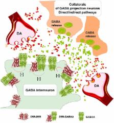
          This perspective article provides observations supporting the view that nigro-striatal dopamine neurons and meso-limbic dopamine neurons mainly communicate through short distance volume transmission in the um range with dopamine diffusing into extrasynaptic and synaptic regions of glutamate and GABA synapses. Based on this communication it is discussed how volume transmission modulates synaptic glutamate transmission onto the D1R modulated direct and D2R modulated indirect GABA pathways of the dorsal striatum. Each nigro-striatal dopamine neuron was first calculated to form large numbers of neostriatal DA nerve terminals and then found to give rise to dense axonal arborizations spread over the neostriatum, from which dopamine is released. These neurons can through DA volume transmission directly influence not only the striatal GABA projection neurons but all the striatal cell types in parallel. It includes the GABA nerve cells forming the island-/striosome GABA pathway to the nigral dopamine cells, the striatal cholinergic interneurons and the striatal GABA interneurons. The dopamine modulation of the different striatal nerve cell types involves the five dopamine receptor subtypes, D1R to D5R receptors, and their formation of multiple extrasynaptic and synaptic dopamine homo and heteroreceptor complexes. These features of the nigro-striatal dopamine neuron to modulate in parallel the activity of practically all the striatal nerve cell types in the dorsal striatum, through the dopamine receptor complexes allows us to understand its unique and crucial fine-tuning of movements, which is lost in Parkinson's disease. Integration of striatal dopamine signals with other transmitter systems in the striatum mainly takes place via the receptor-receptor interactions in dopamine heteroreceptor complexes. Such molecular events also participate in the integration of volume transmission and synaptic transmission. Dopamine modulation of the glutamate synapses on the dorsal striato-pallidal GABA pathway involves D2R heteroreceptor complexes such as D2R-NMDAR, A2AR-D2R, and NTSR1-D2R heteroreceptor complexes. The dopamine modulation of glutamate synapses on the striato-entopeduncular/nigral pathway takes place mainly via D1R heteroreceptor complexes such as D1R-NMDAR, A2R-D1R, and D1R-D3R heteroreceptor complexes. Dopamine modulation of the island/striosome compartment of the dorsal striatum projecting to the nigral dopamine cells involve D4R-MOR heteroreceptor complexes. All these receptor-receptor interactions have relevance for Parkinson's disease and its treatment.

          Related collections

          Most cited references221

          • Record: found
          • Abstract: found
          • Article: not found

          The glutamate homeostasis hypothesis of addiction.

          Addiction is associated with neuroplasticity in the corticostriatal brain circuitry that is important for guiding adaptive behaviour. The hierarchy of corticostriatal information processing that normally permits the prefrontal cortex to regulate reinforcement-seeking behaviours is impaired by chronic drug use. A failure of the prefrontal cortex to control drug-seeking behaviours can be linked to an enduring imbalance between synaptic and non-synaptic glutamate, termed glutamate homeostasis. The imbalance in glutamate homeostasis engenders changes in neuroplasticity that impair communication between the prefrontal cortex and the nucleus accumbens. Some of these pathological changes are amenable to new glutamate- and neuroplasticity-based pharmacotherapies for treating addiction.
            Bookmark
            • Record: found
            • Abstract: found
            • Article: not found

            Dopamine release from the locus coeruleus to the dorsal hippocampus promotes spatial learning and memory.

            Dopamine neurotransmission in the dorsal hippocampus is critical for a range of functions from spatial learning and synaptic plasticity to the deficits underlying psychiatric disorders such as attention-deficit hyperactivity disorder. The ventral tegmental area (VTA) is the presumed source of dopamine in the dorsal hippocampus. However, there is a surprising scarcity of VTA dopamine axons in the dorsal hippocampus despite the dense network of dopamine receptors. We have explored this apparent paradox using optogenetic, biochemical, and behavioral approaches and found that dopaminergic axons and subsequent dopamine release in the dorsal hippocampus originate from neurons of the locus coeruleus (LC). Photostimulation of LC axons produced an increase in dopamine release in the dorsal hippocampus as revealed by high-performance liquid chromatography. Furthermore, optogenetically induced release of dopamine from the LC into the dorsal hippocampus enhanced selective attention and spatial object recognition via the dopamine D1/D5 receptor. These results suggest that spatial learning and memory are energized by the release of dopamine in the dorsal hippocampus from noradrenergic neurons of the LC. The present findings are critical for identifying the neural circuits that enable proper attention selection and successful learning and memory.
              Bookmark
              • Record: found
              • Abstract: found
              • Article: not found

              Heterodimerization is required for the formation of a functional GABA(B) receptor.

              GABA (gamma-aminobutyric acid) is the main inhibitory neurotransmitter in the mammalian central nervous system, where it exerts its effects through ionotropic (GABA(A/C)) receptors to produce fast synaptic inhibition and metabotropic (GABA(B)) receptors to produce slow, prolonged inhibitory signals. The gene encoding a GABA(B) receptor (GABA(B)R1) has been cloned; however, when expressed in mammalian cells this receptor is retained as an immature glycoprotein on intracellular membranes and exhibits low affinity for agonists compared with the endogenous receptor on brain membranes. Here we report the cloning of a complementary DNA encoding a new subtype of the GABAB receptor (GABA(B)R2), which we identified by mining expressed-sequence-tag databases. Yeast two-hybrid screening showed that this new GABA(B)R2-receptor subtype forms heterodimers with GABA(B)R1 through an interaction at their intracellular carboxy-terminal tails. Upon expression with GABA(B)R2 in HEK293T cells, GABA(B)R1 is terminally glycosylated and expressed at the cell surface. Co-expression of the two receptors produces a fully functional GABA(B) receptor at the cell surface; this receptor binds GABA with a high affinity equivalent to that of the endogenous brain receptor. These results indicate that, in vivo, functional brain GABA(B) receptors may be heterodimers composed of GABA(B)R1 and GABA(B)R2.
                Bookmark

                Author and article information

                Contributors
                Journal
                Front Synaptic Neurosci
                Front Synaptic Neurosci
                Front. Synaptic Neurosci.
                Frontiers in Synaptic Neuroscience
                Frontiers Media S.A.
                1663-3563
                10 July 2018
                2018
                : 10
                : 20
                Affiliations
                [1] 1Department of Neuroscience, Karolinska Institutet , Stockholm, Sweden
                [2] 2Section of Physiology, Department of Biomolecular Science, University of Urbino , Urbino, Italy
                [3] 3Observatorio Cubano de Neurociencias, Grupo Bohío-Estudio , Yaguajay, Cuba
                [4] 4Instituto de Fisiología Celular, Universidad Nacional Autónoma de México , Mexico City, Mexico
                [5] 5Faculty of Health Sciences, School of Anatomical Sciences, University of the Witwatersrand , Johannesburg, South Africa
                [6] 6Facultad de Medicina, Instituto de Investigación Biomédica de Málaga , Málaga, Spain
                [7] 7Department of Medical Sciences, University of Ferrara , Ferrara, Italy
                [8] 8Department of Biochemistry and Molecular Biomedicine, Faculty of Biomedicine, University of Barcelona , Barcelona, Spain
                [9] 9Laboratory of Drug Addiction Pharmacology, Department of Pharmacology, Institute of Pharmacology, Polish Academy of Sciences , Kraków, Poland
                [10] 10Department of Cell Biology, Faculty of Sciences, University of Málaga , Málaga, Spain
                [11] 11Department of Life Sciences and Biotechnology (SVEB), University of Ferrara , Ferrara, Italy
                [12] 12CiberNed: Centro de Investigación en Red Enfermedades Neurodegenerativas, Instituto de Salud Carlos III , Madrid, Spain
                Author notes

                Edited by: Fang Liu, Centre for Addiction and Mental Health, Canada

                Reviewed by: Louis-Eric Trudeau, Université de Montréal, Canada; Patrizia Ambrogini, Università degli Studi di Urbino Carlo Bo, Italy

                *Correspondence: Kjell Fuxe kjell.fuxe@ 123456ki.se
                Article
                10.3389/fnsyn.2018.00020
                6048293
                30042672
                d2075640-d39b-4bd6-9221-cdc6489ac8aa
                Copyright © 2018 Borroto-Escuela, Perez De La Mora, Manger, Narváez, Beggiato, Crespo-Ramírez, Navarro, Wydra, Díaz-Cabiale, Rivera, Ferraro, Tanganelli, Filip, Franco and Fuxe.

                This is an open-access article distributed under the terms of the Creative Commons Attribution License (CC BY). The use, distribution or reproduction in other forums is permitted, provided the original author(s) and the copyright owner(s) are credited and that the original publication in this journal is cited, in accordance with accepted academic practice. No use, distribution or reproduction is permitted which does not comply with these terms.

                History
                : 14 January 2018
                : 19 June 2018
                Page count
                Figures: 3, Tables: 0, Equations: 0, References: 235, Pages: 24, Words: 21198
                Funding
                Funded by: Hjärnfonden 10.13039/501100003792
                Award ID: FO2016-0302
                Funded by: Vetenskapsrådet 10.13039/501100004359
                Award ID: 348-2014-4396
                Award ID: 04X-715
                Funded by: Dirección General de Asuntos del Personal Académico, Universidad Nacional Autónoma de México 10.13039/501100006087
                Award ID: IN205217
                Funded by: Consejo Nacional de Ciencia y Tecnología 10.13039/501100007350
                Award ID: 213-01-220173
                Categories
                Neuroscience
                Review

                Neurosciences
                g protein-coupled receptor,dopamine receptor,heteroreceptor complexes,oligomerization,parkinson's diseases,volume transmission,neural plasticity

                Comments

                Comment on this article