28
views
0
recommends
+1 Recommend
0 collections
    0
    shares
      • Record: found
      • Abstract: found
      • Article: not found

      sFRP2 in the aged microenvironment drives melanoma metastasis and therapy resistance

      research-article
      1 , 2 , 1 , 1 , 1 , 1 , 1 , 1 , 1 , 1 , 3 , 1 , 4 , 4 , 4 , 1 , 5 , 1 , 1 , 4 , 4 , 4 , 6 , 6 , 7 , 7 , 7 , 7 , 1 , 1 , 8 , 1 , 8 , 8 , 9 , 10 , 11 , 12 , 12 , 13 , 13 , 14 , 14 , 1 , 5 , 10 , 1 , **
      Nature

      Read this article at

      ScienceOpenPublisherPMC
      Bookmark
          There is no author summary for this article yet. Authors can add summaries to their articles on ScienceOpen to make them more accessible to a non-specialist audience.

          Abstract

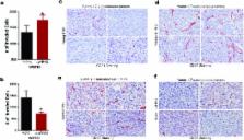
          Cancer is a disease of aging, and aged cancer patients have a poorer prognosis. This may be due to accumulated cellular damage, decreases in adaptive immunity, and chronic inflammation. However, the effects of the aged microenvironment on tumor progression have been largely unexplored. Since dermal fibroblasts can have profound impacts on melanoma progression 14 we examined whether age-related changes in dermal fibroblasts could drive melanoma metastasis and response to targeted therapy. We find that aged fibroblasts secrete a Wnt antagonist, sFRP2, which activates a multi-step signaling cascade in melanoma cells that results in a decrease in β-catenin and MITF, and ultimately the loss of a key redox effector, APE1. Loss of APE1 attenuates the response of melanoma cells to ROS-induced DNA damage, rendering them more resistant to targeted therapy (vemurafenib). Age-related increases in sFRP2 also augment both angiogenesis and metastasis of melanoma cells. These data provide an integrated view of how fibroblasts in the aged microenvironment contribute to tumor progression, offering new paradigms for the design of therapy for the elderly.

          Related collections

          Most cited references22

          • Record: found
          • Abstract: found
          • Article: not found

          Ultraviolet-radiation-induced inflammation promotes angiotropism and metastasis in melanoma.

          Intermittent intense ultraviolet (UV) exposure represents an important aetiological factor in the development of malignant melanoma. The ability of UV radiation to cause tumour-initiating DNA mutations in melanocytes is now firmly established, but how the microenvironmental effects of UV radiation influence melanoma pathogenesis is not fully understood. Here we report that repetitive UV exposure of primary cutaneous melanomas in a genetically engineered mouse model promotes metastatic progression, independent of its tumour-initiating effects. UV irradiation enhanced the expansion of tumour cells along abluminal blood vessel surfaces and increased the number of lung metastases. This effect depended on the recruitment and activation of neutrophils, initiated by the release of high mobility group box 1 (HMGB1) from UV-damaged epidermal keratinocytes and driven by Toll-like receptor 4 (TLR4). The UV-induced neutrophilic inflammatory response stimulated angiogenesis and promoted the ability of melanoma cells to migrate towards endothelial cells and use selective motility cues on their surfaces. Our results not only reveal how UV irradiation of epidermal keratinocytes is sensed by the innate immune system, but also show that the resulting inflammatory response catalyses reciprocal melanoma-endothelial cell interactions leading to perivascular invasion, a phenomenon originally described as angiotropism in human melanomas by histopathologists. Angiotropism represents a hitherto underappreciated mechanism of metastasis that also increases the likelihood of intravasation and haematogenous dissemination. Consistent with our findings, ulcerated primary human melanomas with abundant neutrophils and reactive angiogenesis frequently show angiotropism and a high risk for metastases. Our work indicates that targeting the inflammation-induced phenotypic plasticity of melanoma cells and their association with endothelial cells represent rational strategies to specifically interfere with metastatic progression.
            Bookmark
            • Record: found
            • Abstract: found
            • Article: not found

            Aging and epigenetic drift: a vicious cycle.

            The term epigenetics refers to stable patterns of gene expression that are seen during differentiation or X chromosome inactivation and are not dependent on dynamic changes in coding DNA. These gene expression states are encoded in the epigenome - a collection of marks on DNA or on histone tails that are established during embryogenesis. Genome-wide studies in aging cells and tissues have uncovered stochastic DNA methylation drift (gradual increases or decreases at specific loci) that reflects imperfect maintenance of epigenetic marks. Drift creates epigenetic mosaicism in aging stem cells that could potentially restrict their plasticity and worsen phenotypes such as stem cell exhaustion and focal proliferative defects that can lead to cancer.
              Bookmark
              • Record: found
              • Abstract: found
              • Article: not found

              Ultraviolet radiation accelerates BRAF-driven melanomagenesis by targeting TP53

              Cutaneous melanoma is epidemiologically linked to ultraviolet radiation (UVR), but the molecular mechanisms by which UVR drives melanomagenesis remain unclear 1,2 . The most common somatic mutation in melanoma is a V600E substitution in BRAF, which is an early event 3 . To investigate how UVR accelerates oncogenic BRAF-driven melanomagenesis, we used a V600EBRAF mouse model. In mice expressing V600EBRAF in their melanocytes, a single dose of UVR that mimicked mild sunburn in humans induced clonal expansion of the melanocytes, and repeated doses of UVR increased melanoma burden. We show that sunscreen (UVA superior: UVB SPF50) delayed the onset of UVR-driven melanoma, but only provided partial protection. The UVR-exposed tumours presented increased numbers of single nucleotide variants (SNVs) and we observed mutations (H39Y, S124F, R245C, R270C, C272G) in the Trp53 tumour suppressor in ~40% of cases. TP53 is an accepted UVR target in non-melanoma skin cancer, but is not thought to play a major role in melanoma 4 . However, we show that mutant Trp53 accelerated V600EBRAF-driven melanomagenesis and that TP53 mutations are linked to evidence of UVR-induced DNA damage in human melanoma. Thus, we provide mechanistic insight into epidemiological data linking UVR to acquired naevi in humans 5 . We identify TP53/Trp53 as a UVR-target gene that cooperates with V600EBRAF to induce melanoma, providing molecular insight into how UVR accelerates melanomagenesis. Our study validates public health campaigns that promote sunscreen protection for individuals at risk of melanoma.
                Bookmark

                Author and article information

                Journal
                0410462
                6011
                Nature
                Nature
                Nature
                0028-0836
                1476-4687
                9 February 2016
                04 April 2016
                14 April 2016
                04 October 2016
                : 532
                : 7598
                : 250-254
                Affiliations
                [1 ]The Wistar Institute, Philadelphia, Pennsylvania
                [2 ]University of the Sciences, Philadelphia, Pennsylvania
                [3 ]University of Zurich, Zurich, Switzerland
                [4 ]The National Institute on Aged, NIH, Baltimore, Maryland
                [5 ]Yale University, New Haven, Connecticut
                [6 ]Massachusetts General Hospital Cancer Center, Boston, Massachusetts
                [7 ]MD Anderson Cancer Center, Houston, TX
                [8 ]Abramson Cancer Center, University of Pennsylvania, Philadelphia, Pennsylvania
                [9 ]Department of Medical Oncology, City of Hope Medical Center, Duarte, CA
                [10 ]Department of Medicine, Division of Hematology-Oncology, University of California Los Angeles, Los Angeles, California (UCLA)
                [11 ]Crown Princess Mary Cancer Centre, Westmead Hospital, Westmead, Australia
                [12 ]Melanoma Institute Australia and The University of Sydney, Sydney, Australia
                [13 ]Vanderbilt University Medical Center, Nashville TN
                [14 ]Department of Dermatology, University Hospital, West German Cancer Center, University Duesburg-Essen, Essen, Germany and German Cancer Consortium (DKTK), Heidelberg, Germany
                Author notes
                [** ] To Whom Correspondence Should Be Addressed: Ashani T. Weeraratna, Ph.D., The Wistar Institute, Rm 452/454A, 3601 Spruce Street, Philadelphia, PA 19104, Office: 215 495-6937, Fax: 215 495-6938, aweeraratna@ 123456wistar.org
                Article
                NIHMS757165
                10.1038/nature17392
                4833579
                27042933
                d477317e-908a-486d-806d-0bafe4220b93

                Users may view, print, copy, and download text and data-mine the content in such documents, for the purposes of academic research, subject always to the full Conditions of use: http://www.nature.com/authors/editorial_policies/license.html#terms

                Reprints: Reprints and permissions information is available at www.nature.com/reprints

                History
                Categories
                Article

                Uncategorized
                Uncategorized

                Comments

                Comment on this article