1
views
0
recommends
+1 Recommend
0 collections
    0
    shares
      • Record: found
      • Abstract: found
      • Article: found
      Is Open Access

      A new design for a green calcium indicator with a smaller size and a reduced number of calcium-binding sites

      research-article

      Read this article at

      Bookmark
          There is no author summary for this article yet. Authors can add summaries to their articles on ScienceOpen to make them more accessible to a non-specialist audience.

          Abstract

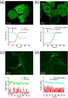
          Genetically encoded calcium indicators (GECIs) are mainly represented by two- or one-fluorophore-based sensors. One type of two-fluorophore-based sensor, carrying Opsanus troponin C (TnC) as the Ca 2+-binding moiety, has two binding sites for calcium ions, providing a linear response to calcium ions. One-fluorophore-based sensors have four Ca 2+-binding sites but are better suited for in vivo experiments. Herein, we describe a novel design for a one-fluorophore-based GECI with two Ca 2+-binding sites. The engineered sensor, called NTnC, uses TnC as the Ca 2+-binding moiety, inserted in the mNeonGreen fluorescent protein. Monomeric NTnC has higher brightness and pH-stability in vitro compared with the standard GECI GCaMP6s. In addition, NTnC shows an inverted fluorescence response to Ca 2+. Using NTnC, we have visualized Ca 2+ dynamics during spontaneous activity of neuronal cultures as confirmed by control NTnC and its mutant, in which the affinity to Ca 2+ is eliminated. Using whole-cell patch clamp, we have demonstrated that NTnC dynamics in neurons are similar to those of GCaMP6s and allow robust detection of single action potentials. Finally, we have used NTnC to visualize Ca 2+ neuronal activity in vivo in the V1 cortical area in awake and freely moving mice using two-photon microscopy or an nVista miniaturized microscope.

          Related collections

          Most cited references14

          • Record: found
          • Abstract: found
          • Article: not found

          Fluorescent indicators for Ca2+ based on green fluorescent proteins and calmodulin.

          Important Ca2+ signals in the cytosol and organelles are often extremely localized and hard to measure. To overcome this problem we have constructed new fluorescent indicators for Ca2+ that are genetically encoded without cofactors and are targetable to specific intracellular locations. We have dubbed these fluorescent indicators 'cameleons'. They consist of tandem fusions of a blue- or cyan-emitting mutant of the green fluorescent protein (GFP), calmodulin, the calmodulin-binding peptide M13, and an enhanced green- or yellow-emitting GFP. Binding of Ca2+ makes calmodulin wrap around the M13 domain, increasing the fluorescence resonance energy transfer (FRET) between the flanking GFPs. Calmodulin mutations can tune the Ca2+ affinities to measure free Ca2+ concentrations in the range 10(-8) to 10(-2) M. We have visualized free Ca2+ dynamics in the cytosol, nucleus and endoplasmic reticulum of single HeLa cells transfected with complementary DNAs encoding chimaeras bearing appropriate localization signals. Ca2+ concentration in the endoplasmic reticulum of individual cells ranged from 60 to 400 microM at rest, and 1 to 50 microM after Ca2+ mobilization. FRET is also an indicator of the reversible intermolecular association of cyan-GFP-labelled calmodulin with yellow-GFP-labelled M13. Thus FRET between GFP mutants can monitor localized Ca2+ signals and protein heterodimerization in individual live cells.
            Bookmark
            • Record: found
            • Abstract: found
            • Article: not found

            A bright monomeric green fluorescent protein derived from Branchiostoma lanceolatum

            Despite the existence of fluorescent proteins spanning the entire visual spectrum, the bulk of modern imaging experiments continue to rely on variants of the green fluorescent protein derived from Aequorea victoria. Meanwhile, a great deal of recent effort has been devoted to engineering and improving red fluorescent proteins, and relatively little attention has been given to green and yellow variants. Here we report a novel monomeric yellow-green fluorescent protein, mNeonGreen, which is derived from a tetrameric fluorescent protein from the cephalochordate Branchiostoma lanceolatum. This fluorescent protein is the brightest monomeric green or yellow fluorescent protein yet described, performs exceptionally well as a fusion tag for traditional imaging as well as stochastic single-molecule superresolution imaging, and is an excellent FRET acceptor for the newest generation of cyan fluorescent proteins.
              Bookmark
              • Record: found
              • Abstract: found
              • Article: not found

              New calcium indicators and buffers with high selectivity against magnesium and protons: design, synthesis, and properties of prototype structures.

              R Tsien (1980)
              A new family of high-affinity buffers and optical indicators for Ca2+ is rationally designed and synthesized. The parent compound is 1,2-bis(o-aminophenoxy)ethane-N,N,N',N'-tetraacetic acid (BAPTA), a relative of the well-known chelator EGTA [ethylene glycol bis(beta-aminoethyl ether)-N,N,N',N'-tetraacetic acid] in which methylene links between oxygen and nitrogen are replaced by benzene rings. BAPTA and its derivatives share the high (greater than 10(5)) selectivity for Ca2+ over Mg2+ of EGTA but are very much less affected by pH changes and are faster at taking up and releasing Ca2+. The affinity of the parent compound for Ca2+ (dissociation constant 1.1 x 10(-7) M in 0.1 M KCl) may be strengthened or weakened by electron-releasing or -withdrawing substituents on the aromatic rings. The Ca2+ and Mg2+ affinities may further be altered by replacing the ether oxygens by heterocyclic nitrogen atoms. The compounds described are fluorescent Ca2+ indicators absorbing in the ultraviolet region; the very large spectral shifts observed on binding Ca2+ fit the prediction that complexation should hinder the conjugation of the nitrogen lone-pair electrons with the aromatic rings. Derivatives with quinoline nuclei are notable for their high sensitivity of fluorescent quantum yield to the binding of Ca2+ but not of Mg2+. Preliminary biological tests have so far revealed little or no binding to membranes or toxic effects following intracellular microinjection.
                Bookmark

                Author and article information

                Journal
                Sci Rep
                Sci Rep
                Scientific Reports
                Nature Publishing Group
                2045-2322
                28 September 2016
                2016
                : 6
                : 34447
                Affiliations
                [1 ]NBICS Department, Moscow Institute of Physics and Technology , Moscow 123182, Russia
                [2 ]Department of Systems Neurobiology and Functional Neurochemistry, P.K. Anokhin Institute of Normal Physiology of RAMS , Moscow 125009, Russia
                [3 ]National Research Center “Kurchatov Institute” , Moscow 123182, Russia
                [4 ]Laboratory of Cellular Neurobiology of Learning, Institute of Higher Nervous Activity and Neurophysiology of RAS , Moscow 117485, Russia
                [5 ]Medico-Biological Faculty, N.I. Pirogov Russian National Research Medical University , Moscow 117997, Russia
                [6 ]MIT Media Lab, Massachusetts Institute of Technology , Cambridge, MA 02139, USA
                [7 ]Federal Research and Clinical Center of Physical-Chemical Medicine of Federal Medical Biological Agency , Moscow 119435, Russia
                [8 ]Engelhardt Institute of Molecular Biology RAS , Moscow 119991, Russia
                [9 ]Cold Spring Harbor Laboratory, Cold Spring Harbor , NY 11724, USA
                [10 ]Department of Anesthesiology, Stony Brook University Medical Center , NY 11794, USA
                [11 ]Center for Developmental Genetics, Stony Brook University , NY 11794, USA
                Author notes
                Article
                srep34447
                10.1038/srep34447
                5039633
                27677952
                d503634b-040a-4c15-9f0f-2e1f55c2863e
                Copyright © 2016, The Author(s)

                This work is licensed under a Creative Commons Attribution 4.0 International License. The images or other third party material in this article are included in the article’s Creative Commons license, unless indicated otherwise in the credit line; if the material is not included under the Creative Commons license, users will need to obtain permission from the license holder to reproduce the material. To view a copy of this license, visit http://creativecommons.org/licenses/by/4.0/

                History
                : 19 May 2016
                : 14 September 2016
                Categories
                Article

                Uncategorized
                Uncategorized

                Comments

                Comment on this article