39
views
0
recommends
+1 Recommend
0 collections
    0
    shares
      • Record: found
      • Abstract: found
      • Article: found
      Is Open Access

      Variations in local calcium signaling in adjacent cardiac myocytes of the intact mouse heart detected with two-dimensional confocal microscopy

      research-article

      Read this article at

      Bookmark
          There is no author summary for this article yet. Authors can add summaries to their articles on ScienceOpen to make them more accessible to a non-specialist audience.

          Abstract

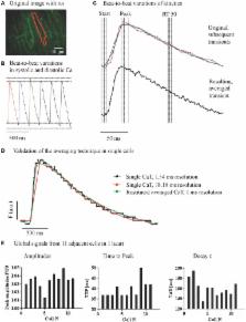
          Dyssynchronous local Ca release within individual cardiac myocytes has been linked to cellular contractile dysfunction. Differences in Ca kinetics in adjacent cells may also provide a substrate for inefficient contraction and arrhythmias. In a new approach we quantify variation in local Ca transients between adjacent myocytes in the whole heart. Langendorff-perfused mouse hearts were loaded with Fluo-8 AM to detect Ca and Di-4-ANEPPS to visualize cell membranes. A spinning disc confocal microscope with a fast camera allowed us to record Ca signals within an area of 465 μm by 315 μm with an acquisition speed of 55 fps. Images from multiple transients recorded at steady state were registered to their time point in the cardiac cycle to restore averaged local Ca transients with a higher temporal resolution. Local Ca transients within and between adjacent myocytes were compared with regard to amplitude, time to peak and decay at steady state stimulation (250 ms cycle length). Image registration from multiple sequential Ca transients allowed reconstruction of high temporal resolution (2.4 ± 1.3 ms) local CaT in 2D image sets ( N = 4 hearts, n = 8 regions). During steady state stimulation, spatial Ca gradients were homogeneous within cells in both directions and independent of distance between measured points. Variation in CaT amplitudes was similar across the short and the long side of neighboring cells. Variations in TAU and TTP were similar in both directions. Isoproterenol enhanced the CaT but not the overall pattern of spatial heterogeneities. Here we detected and analyzed local Ca signals in intact mouse hearts with high temporal and spatial resolution, taking into account 2D arrangement of the cells. We observed significant differences in the variation of CaT amplitude along the long and short axis of cardiac myocytes. Variations of Ca signals between neighboring cells may contribute to the substrate of cardiac remodeling.

          Related collections

          Most cited references34

          • Record: found
          • Abstract: found
          • Article: found
          Is Open Access

          Remodelling of gap junctions and connexin expression in diseased myocardium

          Gap junctions form the cell-to-cell pathways for propagation of the precisely orchestrated patterns of current flow that govern the regular rhythm of the healthy heart. As in most tissues and organs, multiple connexin types are expressed in the heart: connexin43 (Cx43), Cx40 and Cx45 are found in distinctive combinations and relative quantities in different, functionally-specialized subsets of cardiac myocyte. Mutations in genes that encode connexins have only rarely been identified as being a cause of human cardiac disease, but remodelling of connexin expression and gap junction organization are well documented in acquired adult heart disease, notably ischaemic heart disease and heart failure. Remodelling may take the form of alterations in (i) the distribution of gap junctions and (ii) the amount and type of connexins expressed. Heterogeneous reduction in Cx43 expression and disordering in gap junction distribution feature in human ventricular disease and correlate with electrophysiologically identified arrhythmic changes and contractile dysfunction in animal models. Disease-related alterations in Cx45 and Cx40 expression have also been reported, and some of the functional implications of these are beginning to emerge. Apart from ventricular disease, various features of gap junction organization and connexin expression have been implicated in the initiation and persistence of the most common form of atrial arrhythmia, atrial fibrillation, though the disparate findings in this area remain to be clarified. Other major tasks ahead focus on the Purkinje/working ventricular myocyte interface and its role in normal and abnormal impulse propagation, connexin-interacting proteins and their regulatory functions, and on defining the precise functional properties conferred by the distinctive connexin co-expression patterns of different myocyte types in health and disease.
            Bookmark
            • Record: found
            • Abstract: found
            • Article: not found

            So little source, so much sink: requirements for afterdepolarizations to propagate in tissue.

            How early (EADs) and delayed afterdepolarizations (DADs) overcome electrotonic source-sink mismatches in tissue to trigger premature ventricular complexes remains incompletely understood. To study this question, we used a rabbit ventricular action potential model to simulate tissues in which a central area of contiguous myocytes susceptible to EADs or DADs was surrounded by unsusceptible tissue. In 1D tissue with normal longitudinal conduction velocity (0.55 m/s), the numbers of contiguous susceptible myocytes required for an EAD and a barely suprathreshold DAD to trigger a propagating action potential were 70 and 80, respectively. In 2D tissue, these numbers increased to 6940 and 7854, and in 3D tissue to 696,910 and 817,280. These numbers were significantly decreased by reduced gap junction conductance, simulated fibrosis, reduced repolarization reserve and heart failure electrical remodeling. In conclusion, the source-sink mismatch in well-coupled cardiac tissue powerfully protects the heart from arrhythmias due to sporadic afterdepolarizations. Structural and electrophysiological remodeling decrease these numbers significantly but still require synchronization mechanisms for EADs and DADs to overcome the robust protective effects of source-sink mismatch. Copyright 2010 Biophysical Society. Published by Elsevier Inc. All rights reserved.
              Bookmark
              • Record: found
              • Abstract: found
              • Article: not found

              Reduced synchrony of Ca2+ release with loss of T-tubules-a comparison to Ca2+ release in human failing cardiomyocytes.

              During cardiac excitation-contraction coupling, Ca2+ release from the sarcoplasmic reticulum (SR) occurs at the junctional complex with the T-tubules, containing the L-type Ca2+ channels. A partial loss of T-tubules has been described in myocytes from failing canine and human hearts. We examined how graded reduction of T-tubule density would affect the synchrony of Ca2+ release. Adult pig ventricular myocytes were isolated and cultured for 24 and 72 h. T-tubules, visualized with di-8-ANEPPS, and [Ca2+]i transients (Fluo-3) were recorded during confocal line scan imaging. Cultured cardiomyocytes exhibited a progressive reduction in T-tubule density. [Ca2+]i transients showed small areas of delayed Ca2+ release which gradually increased in number and size with loss of T-tubules. Local [Ca2+]i transients in the delayed regions were reduced. Due to these changes, loss of T-tubules resulted in an overall slowing of the rise of [Ca2+] along the entire line scan and transient magnitude tended to be reduced, but there was no change in SR Ca2+ content. Human myocytes isolated from failing hearts had a T-tubule density comparable to that of freshly isolated pig myocytes. The size, but not the number, of delayed release areas tended to be larger. The overall rate of rise of [Ca2+]i was significantly faster than in pig myocytes with low T-tubule density. Loss of T-tubules reduces the synchrony of SR Ca2+ release. This could contribute to reduced efficiency of excitation-contraction coupling in heart failure, though dyssynchrony in human failing cells appears to be modest.
                Bookmark

                Author and article information

                Contributors
                Journal
                Front Physiol
                Front Physiol
                Front. Physiol.
                Frontiers in Physiology
                Frontiers Media S.A.
                1664-042X
                12 January 2015
                2014
                : 5
                : 517
                Affiliations
                [1] 1Department of Cardiology, Medical University of Graz Graz, Austria
                [2] 2Department of Internal Medicine II, University Hospital Regensburg Regensburg, Germany
                [3] 3Molecular Biophysics and Physiology, Rush Medical College, Rush University Chicago, IL, USA
                [4] 4Department of Cardiology, Charité-Universitaetsmedizin Berlin Berlin, Germany
                Author notes

                Edited by: Gil Bub, University of Oxford, UK

                Reviewed by: Andrew F. James, University of Bristol, UK; Cynthia Carnes, The Ohio State University, USA; Antonius Baartscheer, Academic Medical Center, Netherlands

                *Correspondence: Frank R. Heinzel, Department of Cardiology, Charité-Universitaetsmedizin Berlin, Charité Campus Virchow Klinikum, Augustenburger Platz 1, 13353 Berlin, Germany e-mail: frank.heinzel@ 123456medunigraz.at

                This article was submitted to Cardiac Electrophysiology, a section of the journal Frontiers in Physiology.

                Article
                10.3389/fphys.2014.00517
                4290493
                25628569
                d5980fad-1c62-4088-9ea7-2c3c0c17db37
                Copyright © 2015 Hammer, Hohendanner, Blatter, Pieske and Heinzel.

                This is an open-access article distributed under the terms of the Creative Commons Attribution License (CC BY). The use, distribution or reproduction in other forums is permitted, provided the original author(s) or licensor are credited and that the original publication in this journal is cited, in accordance with accepted academic practice. No use, distribution or reproduction is permitted which does not comply with these terms.

                History
                : 27 August 2014
                : 18 December 2014
                Page count
                Figures: 6, Tables: 0, Equations: 0, References: 44, Pages: 11, Words: 7821
                Categories
                Physiology
                Original Research Article

                Anatomy & Physiology
                whole heart,calcium cycling,excitation contraction coupling,2d confocal microscopy,langendorff perfused heart,cardiac myocytes,local dyssynchrony,intercellular communication

                Comments

                Comment on this article