12
views
0
recommends
+1 Recommend
0 collections
    0
    shares
      • Record: found
      • Abstract: found
      • Article: found
      Is Open Access

      The Activity of KIF14, Mieap, and EZR in a New Type of the Invasive Component, Torpedo-Like Structures, Predetermines the Metastatic Potential of Breast Cancer

      research-article

      Read this article at

      Bookmark
          There is no author summary for this article yet. Authors can add summaries to their articles on ScienceOpen to make them more accessible to a non-specialist audience.

          Abstract

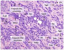
          Intratumor morphological heterogeneity reflects patterns of invasive growth and is an indicator of the metastatic potential of breast cancer. In this study, we used this heterogeneity to identify molecules associated with breast cancer invasion and metastasis. The gene expression microarray data were used to identify genes differentially expressed between solid, trabecular, and other morphological arrangements of tumor cells. Immunohistochemistry was applied to evaluate the association of the selected proteins with metastasis. RNA-sequencing was performed to analyze the molecular makeup of metastatic tumor cells. High frequency of metastases and decreased metastasis-free survival were detected in patients either with positive expression of KIF14 or Mieap or negative expression of EZR at the tips of the torpedo-like structures in breast cancers. KIF14- and Mieap-positive and EZR-negative cells were mainly detected in the torpedo-like structures of the same breast tumors; however, their transcriptomic features differed. KIF14-positive cells showed a significant upregulation of genes involved in ether lipid metabolism. Mieap-positive cells were enriched in genes involved in mitophagy. EZR-negative cells displayed upregulated genes associated with phagocytosis and the chemokine-mediated signaling pathway. In conclusion, the positive expression of KIF14 and Mieap and negative expression of EZR at the tips of the torpedo-like structures are associated with breast cancer metastasis.

          Related collections

          Most cited references36

          • Record: found
          • Abstract: found
          • Article: not found

          Analysis of the kinesin superfamily: insights into structure and function.

          Kinesin superfamily proteins (KIFs) are key players or 'hub' proteins in the intracellular transport system, which is essential for cellular function and morphology. The KIF superfamily is also the first large protein family in mammals whose constituents have been completely identified and confirmed both in silico and in vivo. Numerous studies have revealed the structures and functions of individual family members; however, the relationships between members or a perspective of the whole superfamily structure until recently remained elusive. Here, we present a comprehensive summary based on a large, systematic phylogenetic analysis of the kinesin superfamily. All available sequences in public databases, including genomic information from all model organisms, were analyzed to yield the most complete phylogenetic kinesin tree thus far, comprising 14 families. This comprehensive classification builds on the recently proposed standardized nomenclature for kinesins and allows systematic analysis of the structural and functional relationships within the kinesin superfamily.
            Bookmark
            • Record: found
            • Abstract: found
            • Article: not found

            Lung metastasis genes couple breast tumor size and metastatic spread.

            The association between large tumor size and metastatic risk in a majority of clinical cancers has led to questions as to whether these observations are causally related or whether one is simply a marker for the other. This is partly due to an uncertainty about how metastasis-promoting gene expression changes can arise in primary tumors. We investigated this question through the analysis of a previously defined "lung metastasis gene-expression signature" (LMS) that mediates experimental breast cancer metastasis selectively to the lung and is expressed by primary human breast cancer with a high risk for developing lung metastasis. Experimentally, we demonstrate that the LMS promotes primary tumor growth that enriches for LMS(+) cells, and it allows for intravasation after reaching a critical tumor size. Clinically, this corresponds to LMS(+) tumors being larger at diagnosis compared with LMS(-) tumors and to a marked rise in the incidence of metastasis after LMS(+) tumors reach 2 cm. Patients with LMS-expressing primary tumors selectively fail in the lung compared with the bone or other visceral sites and have a worse overall survival. The mechanistic linkage between metastasis gene expression, accelerated tumor growth, and likelihood of metastatic recurrence provided by the LMS may help to explain observations of prognostic gene signatures in primary cancer and how tumor growth can both lead to metastasis and be a marker for cells destined to metastasize.
              Bookmark
              • Record: found
              • Abstract: found
              • Article: not found

              KIF14 and citron kinase act together to promote efficient cytokinesis

              Multiple mitotic kinesins and microtubule-associated proteins (MAPs) act in concert to direct cytokinesis (Glotzer, M. 2005. Science. 307:1735–1739). In anaphase cells, many of these proteins associate with an antiparallel array of microtubules termed the central spindle. The MAP and microtubule-bundling protein PRC1 (protein-regulating cytokinesis 1) is one of the key molecules required for the integrity of this structure (Jiang, W., G. Jimenez, N.J. Wells, T.J. Hope, G.M. Wahl, T. Hunter, and R. Fukunaga. 1998. Mol. Cell. 2:877–885; Mollinari, C., J.P. Kleman, W. Jiang, G. Schoehn, T. Hunter, and R.L. Margolis. 2002. J. Cell Biol. 157:1175–1186). In this study, we identify an interaction between endogenous PRC1 and the previously uncharacterized kinesin KIF14 as well as other mitotic kinesins (MKlp1/CHO1, MKlp2, and KIF4) with known functions in cytokinesis (Hill, E., M. Clarke, and F.A. Barr. 2000. EMBO J. 19:5711–5719; Matuliene, J., and R. Kuriyama. 2002. Mol. Biol. Cell. 13:1832–1845; Kurasawa, Y., W.C. Earnshaw, Y. Mochizuki, N. Dohmae, and K. Todokoro. 2004. EMBO J. 23:3237–3248). We find that KIF14 targets to the central spindle via its interaction with PRC1 and has an essential function in cytokinesis. In KIF14-depleted cells, citron kinase but not other components of the central spindle and cleavage furrow fail to localize. Furthermore, the localization of KIF14 and citron kinase to the central spindle and midbody is codependent, and they form a complex depending on the activation state of citron kinase. Contrary to a previous study (Di Cunto, F., S. Imarisio, E. Hirsch, V. Broccoli, A. Bulfone, A. Migheli, C. Atzori, E. Turco, R. Triolo, G.P. Dotto, et al. 2000. Neuron. 28:115–127), we find a general requirement for citron kinase in human cell division. Together, these findings identify a novel pathway required for efficient cytokinesis.
                Bookmark

                Author and article information

                Journal
                Cancers (Basel)
                Cancers (Basel)
                cancers
                Cancers
                MDPI
                2072-6694
                15 July 2020
                July 2020
                : 12
                : 7
                : 1909
                Affiliations
                [1 ]Laboratory of Cancer Progression Biology, Cancer Research Institute, Tomsk National Research Medical Center, Russian Academy of Sciences, 634009 Tomsk, Russia; t_gerashchenko@ 123456oncology.tomsk.ru (T.S.G.); zolotarevasyu@ 123456oncology.tomsk.ru (S.Y.Z.); kiselev_am@ 123456almazovcentre.ru (A.M.K.); novikovnm@ 123456oncology.tomsk.ru (N.M.N.)
                [2 ]Institute of Cytology, Russian Academy of Sciences, 194064 Saint Petersburg, Russia
                [3 ]Department of General and Molecular Pathology, Cancer Research Institute, Tomsk National Research Medical Center, Russian Academy of Sciences, 634009 Tomsk, Russia; tashireva@ 123456oncology.tomsk.ru (L.A.T.); pk_ssmu@ 123456ssmu.ru (M.V.Z.); pvm@ 123456ngs.ru (V.M.P.)
                [4 ]Department of Pathological Anatomy, Siberian State Medical University, 634050 Tomsk, Russia; kaf.pat.anatom@ 123456ssmu.ru
                [5 ]Laboratory of Molecular Oncology and Immunology, Cancer Research Institute, Tomsk National Research Medical Center, Russian Academy of Sciences, 634009 Tomsk, Russia; nvch@ 123456tnimc.ru
                Author notes
                [* ]Correspondence: d_evgeniy@ 123456oncology.tomsk.ru ; Tel.: +7-3822-282676
                Author information
                https://orcid.org/0000-0002-7283-0092
                https://orcid.org/0000-0003-2061-8417
                https://orcid.org/0000-0003-2923-9755
                Article
                cancers-12-01909
                10.3390/cancers12071909
                7409151
                32679794
                d6e199cf-13ad-4cbb-8ae3-56cb00098436
                © 2020 by the authors.

                Licensee MDPI, Basel, Switzerland. This article is an open access article distributed under the terms and conditions of the Creative Commons Attribution (CC BY) license ( http://creativecommons.org/licenses/by/4.0/).

                History
                : 04 June 2020
                : 13 July 2020
                Categories
                Article

                breast cancer,metastasis,invasion,heterogeneity,morphology,torpedo-like structures

                Comments

                Comment on this article