14
views
0
recommends
+1 Recommend
0 collections
    0
    shares
      • Record: found
      • Abstract: found
      • Article: found
      Is Open Access

      Congenital muscular torticollis

      case-report

      Read this article at

      Bookmark
          There is no author summary for this article yet. Authors can add summaries to their articles on ScienceOpen to make them more accessible to a non-specialist audience.

          Abstract

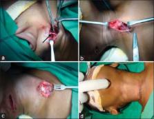
          Congenital muscular torticollis (CMT) is a rare congenital musculoskeletal disorder characterized by unilateral shortening of the sternocleidomastoid muscle (SCM). It presents in newborn infants or young children with reported incidence ranging from 0.3% to 2%. Owing to effective shortening of SCM on the involved side there is ipsilateral head tilt and contralateral rotation of the face and chin. This article reports a case of CMT in a 3½-year-old male child successfully managed by surgical release of the involved SCM followed by physiotherapy.

          Related collections

          Most cited references15

          • Record: found
          • Abstract: found
          • Article: not found

          Congenital muscular torticollis: current concepts and review of treatment.

          Phuong Do (2006)
          The purpose of this review is to better understand the spectrum of disease in torticollis, which is the third most common pediatric orthopaedic diagnosis in childhood. Besides the benign muscular tightness of the sternocleidomastoid muscle leading to the classic head position, the differential diagnosis of the wry neck include sequelae to inflammatory, ocular, neurologic or orthopedic diseases. Patients present with a stiff and tilted neck, and therefore require a thorough and systematic work-up, including a complete physical and neurologic examination and cervical spine radiographs. Recent findings show that magnetic resonance imaging of the brain and neck is no longer considered cost-effective, or necessary, in congenital muscular torticollis. Observation and physical therapy, with or without bracing, is usually an effective treatment in most cases, especially if instituted within the first year of life. Botox has recently been shown to be an effective intermediate method of treatment for more resistant cases of congenital muscular torticollis. In those presenting after the age of 1 year, there is an increased rate of sternocleidomastoid muscle lengthening. The lengthening may improve the range of motion, but not necessarily the plagiocephaly, facial asymmetry, or cranial molding. It is important to differentiate muscular from nonmuscular torticollis. Congenital muscular torticollis is benign; missing a case of nonmuscular torticollis could be potentially life threatening.
            Bookmark
            • Record: found
            • Abstract: found
            • Article: not found

            Infantile torticollis: a review of 624 cases.

            J Cheng, A W Au (2015)
            We reviewed 624 cases of infantile torticollis in one centre over a period of 7 years. The incidence of torticollis was found to be 1.3% in Chinese children. Boy-to-girl ratio was 3:2. Obstetric histories of the mothers showed a total of 62.2% with difficult labour, breech deliveries, or caesarean section, and 6.04% had associated congenital anomalies. Of all the cases, 27.88% were found to be postural, 35.4% had torticollis that presented with sternomastoid tumor, and 36.7% presented with muscular torticollis alone. When limitation of neck range was considered, 36.7% had a passive rotation deficit > 15 degrees. In patients presenting in the early stages, 97% of all infantile torticollis cases resolved with conservative treatment, active stimulation, and a passive stretching program. For those responding to treatment, the mean treatment period was 30 degrees were more likely to need surgery. Presence of sternomastoid tumor alone was not found to increase the likelihood of surgery. Musculoskeletal sequelae after torticollis had resolved included intermittent head tilt and persistence of mild craniofacial asymmetry. We recommend continuous follow-up in cases of infantile torticollis, particularly in patients with progression of sternomastoid tumor to muscular torticollis.
              Bookmark
              • Record: found
              • Abstract: found
              • Article: not found

              Congenital muscular torticollis: sequela of intrauterine or perinatal compartment syndrome.

              The etiology of congenital muscular torticollis remains a mystery despite intensive investigation. Magnetic resonance imaging (MRI) scans of 10 infants with this condition showed signals in the sternocleidomastoid muscle similar to those observed in the forearm and leg after compartment syndrome. Cadaver dissections and injection studies defined the sternocleidomastoid muscle compartment. Injection studies and pressure measurements performed at the time of bipolar release in three patients with congenital muscular torticollis confirmed the existence of this compartment in vivo. Clinical review of 48 children with congenital muscular torticollis showed a relation between birth position and the side affected by the contracture. Because of the association of congenital muscular torticollis with other intrauterine positioning disorders, we postulate that head positioning in utero can selectively injure the sternocleidomastoid muscle, leading to development of a compartment syndrome. Congenital muscular torticollis may represent the sequela of an intrauterine or perinatal compartment syndrome.
                Bookmark

                Author and article information

                Journal
                Ann Maxillofac Surg
                Ann Maxillofac Surg
                AMS
                Annals of Maxillofacial Surgery
                Medknow Publications & Media Pvt Ltd (India )
                2231-0746
                2249-3816
                Jul-Dec 2013
                : 3
                : 2
                : 198-200
                Affiliations
                [1]Department of Oral and Maxillofacial Surgery, School of Dental Sciences, KIMSDU, Karad, Maharashtra, India
                [1 ]Department of Oral and Maxillofacial Surgery, Calcutta Institute of Maxillofacial Surgery and Research, Kolkata, West Bengal, India
                Author notes
                Address for correspondence: Dr. Kumar Nilesh, S/o. Dr. R. P. Mishra, Radha Sadan, Raj Guru Path, Bilasi, Deoghar - 814 117, Jharkhand, India. E-mail: drkumarnilesh@ 123456yahoo.com
                Article
                AMS-3-198
                10.4103/2231-0746.119222
                3814673
                d7cea4bf-a444-4e1b-b963-8944e76efb85
                Copyright: © Annals of Maxillofacial Surgery

                This is an open-access article distributed under the terms of the Creative Commons Attribution-Noncommercial-Share Alike 3.0 Unported, which permits unrestricted use, distribution, and reproduction in any medium, provided the original work is properly cited.

                History
                Categories
                Case Report - Developmental Disturbances

                congenital,sternocleidomastoid muscle,tenotomy,torticollis

                Comments

                Comment on this article