6
views
0
recommends
+1 Recommend
0 collections
    0
    shares
      • Record: found
      • Abstract: found
      • Article: found
      Is Open Access

      Strain Elastography Ultrasound: An Overview with Emphasis on Breast Cancer Diagnosis

      review-article

      Read this article at

      Bookmark
          There is no author summary for this article yet. Authors can add summaries to their articles on ScienceOpen to make them more accessible to a non-specialist audience.

          Abstract

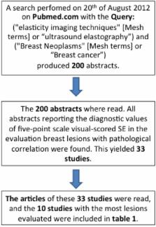
          Strain elastography (SE), which estimates tissue strain, is an adjunct to the conventional ultrasound B-mode examination. We present a short introduction to SE and its clinical use. Furthermore, we present an overview of the 10 largest studies performed on the diagnostic accuracy of SE in breast cancer diagnostics. Eight of 10 studies presented data for both SE and B-mode imaging. Seven studies showed better specificity and accuracy for SE than for B-mode imaging in breast cancer diagnosis. Four studies showed an increase in specificity and accuracy when combining B-mode imaging with SE. The ways of combining B-mode imaging with SE in the diagnosis of breast cancer differed between the five studies. We believe that further studies are needed to establish an optimal algorithm for the combination of B-mode ultrasound and SE in breast cancer.

          Related collections

          Most cited references28

          • Record: found
          • Abstract: found
          • Article: not found

          Elastography of breast lesions: initial clinical results.

          To determine the appearance of various breast lesions on elastograms and to explore the potential of elastography in the diagnosis of breast lesions. A total of 46 breast lesions were examined with elastography. Patients underwent biopsy or aspiration of all lesions, revealing 15 fibroadenomas, 12 carcinomas, six fibrocystic nodules, and 13 other lesions. The elastogram was generated from radio-frequency data collected with use of a 5-MHz linear-array transducer. The elastogram and corresponding sonogram were evaluated by a single observer for lesion visualization, relative brightness, and margin definition and regularity. The sizes of the lesions at each imaging examination and at biopsy were recorded and compared. Softer tissues such as fat appear as bright areas on elastograms. Firm tissues, including parenchyma, cancers, and other masses, appear darker. The cancers were statistically significantly darker than fibroadenomas (P < .005) and substantially larger on the elastogram than on the sonogram. Seventy-three percent of fibroadenomas and 56% of solid benign lesions could be distinguished from cancers by using lesion brightness and size difference. Some cancers that appeared as areas of shadowing on sonograms appeared as discrete masses on elastograms. Elastography has the potential to be useful in the evaluation of areas of shadowing on the sonogram. It also may be helpful in the distinction of benign from malignant masses.
            Bookmark
            • Record: found
            • Abstract: found
            • Article: not found

            Interobserver agreement in assessing the sonographic and elastographic features of malignant thyroid nodules.

            The objective of our study was to investigate interobserver agreement for the diagnosis of malignant thyroid nodules using conventional B-mode ultrasound and real-time freehand ultrasound elastography. Between December 2007 and February 2008, 45 patients (age range, 19-73 years; mean age +/- SD, 45.0 +/- 12.2 years) with 52 thyroid nodules were examined with conventional B-mode ultrasound and real-time freehand ultrasound elastography. All the patients were scheduled to undergo thyroid surgery because a thyroid nodule had been proven malignant on aspiration cytology. Three radiologists independently performed conventional ultrasound and elastography and analyzed the ultrasound images. Using conventional ultrasound, observers recorded the following information about nodular features: composition (solid, cystic, or mixed cystic-solid), echogenicity (hyperechoic, isoechoic, hypoechoic, or markedly hypoechoic), margin (well circumscribed, microlobulated, or irregular), calcification (negative [no calcifications]; microcalcification, macrocalcification, or mixed-type calcifications), and shape (parallel or nonparallel). Observers determined the Ueno classification and area ratio for each nodule using ultrasound elastography. Interobserver agreement was evaluated with Spearman's correlation analysis for all findings except the area ratio, for which Pearson's correlation analysis was used. A p 0.05) or the area ratio (Pearson's correlation coefficient, -0.03 to 0.23; p > 0.05). Statistically significant concordance among radiologists about most features of malignant thyroid nodules was seen with conventional ultrasound; however, ultrasound elastography did not show reliable interobserver agreement for the diagnosis of malignant thyroid nodules.
              Bookmark
              • Record: found
              • Abstract: found
              • Article: not found

              Significant differentiation of focal breast lesions: calculation of strain ratio in breast sonoelastography.

              Initial data suggest that elastography can improve the specificity of ultrasound in differentiating benign and malignant breast lesions. The aim of this study was to compare elastography and B-mode ultrasound to determine whether the calculation of strain ratios (SRs) can further improve the differentiation of focal breast lesions. A total of 227 women with histologically proven focal breast lesions (113 benign, 114 malignant) were included at two German breast centers. The women underwent a standardized ultrasound procedure using a high-end ultrasound system with a 9-MHz broadband linear transducer. B-mode scans and sonoelastograms were analyzed by two experienced readers using the Breast Imaging Reporting and Data System criteria. SRs were calculated from a tumor-adjusted region of interest (mean color pixel density) and a comparable region of interest placed in the lateral fatty tissue. Sensitivity, specificity, and cutoff values were calculated for SRs (receiver-operating characteristic analysis). The women had a mean age of 54 years (range, 19-87 years). The mean lesion diameter was 1.6 +/- 0.9 cm. Sensitivity and specificity were 96% and 56% for B-mode scanning, 81% and 89% for elastography, and 90% and 89% for SRs. An SR cutoff value of 2.45 (area under the curve, 0.949) allowed significant differentiation (P < .001) of malignant (mean, 5.1 +/- 4.2) and benign (mean, 1.6 +/- 1.0) lesions. The quantitative method of SR calculation was superior to subjective interpretation of sonoelastograms and B-mode scans, with a positive predictive value of 89% compared to 68% and 84% for the other two methods. Calculation of SRs contributes to the standardization of sonoelastography with high sensitivity and allows significant differentiation of benign and malignant breast lesions with higher specificity compared to B-mode ultrasound but not elastography. Copyright 2010 AUR. Published by Elsevier Inc. All rights reserved.
                Bookmark

                Author and article information

                Journal
                Diagnostics (Basel)
                Diagnostics (Basel)
                diagnostics
                Diagnostics
                MDPI
                2075-4418
                25 February 2013
                March 2013
                : 3
                : 1
                : 117-125
                Affiliations
                Department of Radiology, Rigshospitalet, Copenhagen University Hospital, Blegdamsvej 9, Copenhagen OE DK-2100, Denmark; E-Mails: caroline.ewertsen@ 123456dadlnet.dk (C.E.); lönn.lars@ 123456gmail.com (L.L.); mbn@ 123456dadlnet.dk (M.B.N.)
                Author notes
                [* ]Author to whom correspondence should be addressed; E-Mail: jonathan.carlsen@ 123456gmail.com ; Tel.: +45-61-270-407; Fax: +45-35-452-058.
                Article
                diagnostics-03-00117
                10.3390/diagnostics3010117
                4665583
                26835671
                d8ffcc67-30ae-408b-abc5-9e6558753011
                © 2013 by the authors; licensee MDPI, Basel, Switzerland.

                This article is an open access article distributed under the terms and conditions of the Creative Commons Attribution license ( http://creativecommons.org/licenses/by/3.0/).

                History
                : 31 December 2012
                : 11 February 2013
                : 20 February 2013
                Categories
                Review

                strain elastography,ultrasound,breast cancer,diagnostic accuracy

                Comments

                Comment on this article