4
views
0
recommends
+1 Recommend
0 collections
    0
    shares
      • Record: found
      • Abstract: found
      • Article: found
      Is Open Access

      Bioceramic hydroxyapatite-based scaffold with a porous structure using honeycomb as a natural polymeric Porogen for bone tissue engineering

      research-article

      Read this article at

      Bookmark
          There is no author summary for this article yet. Authors can add summaries to their articles on ScienceOpen to make them more accessible to a non-specialist audience.

          Abstract

          Background

          The application of bioceramic hydroxyapatite (HA) derived from materials high in calcium to tissue engineering has been of concern, namely scaffold. Scaffold pores allow for cell mobility metabolic processes, and delivery of oxygen and nutrients by blood vessel. Thus, pore architecture affects cell seeding efficiency, cell viability, migration, morphology, cell proliferation, cell differentiation, angiogenesis, mechanical strength of scaffolds, and, eventually, bone formation. Therefore, to improve the efficacy of bone regeneration, several important parameters of the pore architecture of scaffolds must be carefully controlled, including pore size, geometry, orientation, uniformity, interconnectivity, and porosity, which are interrelated and whose coordination affects the effectiveness of bone tissue engineering. The honeycomb (HCB) as natural polymeric porogen is used to pore forming agent of scaffolds. It is unique for fully interconnected and oriented pores of uniform size and high mechanical strength in the direction of the pores. The aim of this study was therefore to evaluate the effect of HCB concentration on macropore structure of the scaffolds.

          Methods

          Bioceramic hydroxyapatite (HA) was synthesized from abalone mussel shells ( Halioitis asinina) using a precipitation method, and HA-based scaffolds were fabricated with honeycomb (HCB) as the porogen agent. Pore structure engineering was successfully carried out using HCB at concentrations of 10, 20, and 30 wt%.

          Results

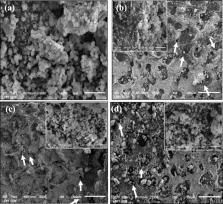
          The Energy Dispersive X-Ray Spectroscopy (EDS) analysis revealed that the Ca/P molar ratio of HA was 1.67 (the stoichiometric ratio of HA). The Fourier Transform Infrared Spectroscopy (FTIR) spectra results for porous HA-based scaffolds and synthesized HA showed that no chemical decomposition occurred in the HA-based scaffold fabrication process. The porosity of the scaffold tended to increase when higher concentrations of HCB were added. XRD data show that the HCB was completely degraded from the scaffold material. The cell metabolic activity and morphology of the HA + HCB 30 wt% scaffold enable it to facilitate the attachment of MC3T3E1 cells on its surface.

          Conclusion

          HCB 30 wt% is the best concentration to fabricate the scaffold corresponding to the criteria for pores structure, crystallographic properties, chemical decomposition process and cell viability for bone tissue engineering.

          Related collections

          Most cited references28

          • Record: found
          • Abstract: found
          • Article: found
          Is Open Access

          Syntheses of hydroxyapatite from natural sources

          Waste materials from natural sources are important resources for extraction and recovery of valuable compounds. Transformation of these waste materials into valuable materials requires specific techniques and approaches. Hydroxyapatite (HAp) is a biomaterial that can be extracted from natural wastes. HAp has been widely used in biomedical applications owing to its excellent bioactivity, high biocompatibility, and excellent osteoconduction characteristics. Thus, HAp is gaining prominence for applications as orthopaedic implants and dental materials. This review summarizes some of the recent methods for extraction of HAp from natural sources including mammalian, aquatic or marine sources, shell sources, plants and algae, and from mineral sources. The extraction methods used to obtain hydroxyapatite are also described. The effect of extraction process and natural waste source on the critical properties of the HAp such as Ca/P ratio, crystallinity and phase assemblage, particle sizes, and morphology are discussed herein.
            Bookmark
            • Record: found
            • Abstract: found
            • Article: not found

            Preparation and histological evaluation of biomimetic three-dimensional hydroxyapatite/chitosan-gelatin network composite scaffolds.

            A novel biodegradable hydroxyapatite/chitosan-gelatin network (HA/CS-Gel) composite of similar composition to that of normal human bone was prepared as a three-dimensional biomimetic scaffold by phase separation method for bone tissue engineering. Changing the solid content and the compositional variables of the original mixtures allowed control of the porosities and densities of the scaffolds. The HA granules were dispersed uniformly in the organic network with intimate interface contact via pulverizing and ultrasonically treating commercial available HA particles. Scaffolds of 90.6% porosity were used to examine the proliferation and functions of the cells in this three-dimensional microenvironment by culturing neonatal rat caldaria osteoblasts. Histological and immunohistochemical staining and scanning electron microscopy observation indicated that the osteoblasts attached to and proliferated on the scaffolds. Extracellular matrices including collagen I and proteoglycan-like substrate were synthesized, while osteoid and bone-like tissue formed during the culture period. Furthermore, the cell/scaffold constructs had good biomineralization effect after 3 weeks in culture.
              Bookmark
              • Record: found
              • Abstract: found
              • Article: not found

              Biomimetic composite scaffold of hydroxyapatite/gelatin-chitosan core-shell nanofibers for bone tissue engineering

              Good biocompatibility and osteogenesis of three-dimensional porous scaffolds are critical for bone tissue engineering. In this work, biomimetic hydroxyapatite/gelatin-chitosan core-shell nanofibers composite scaffolds have been fabricated to mimic both the specific structure and the chemical composition of natural bone. The coaxial electrospinning technique was introduced to prepare gelatin-chitosan core-shell structured nanofibers mat which formed three-dimensional porous structure for promoting cells growth. The gelatin-chitosan core-shell nanofibers formed Arginine-Glycine-Aspartic acid (RGD)-like structure to mimic the organic component of natural bone extracellular matrix. Hydroxyapatite (Ca10(PO4)6(OH)2, HAP), as the major mineral constituent of native bone, was then deposited onto the surface of gelatin-chitosan core-shell structured nanofibers by a wet chemical method. Compared with chitosan nanofibers, gelatin nanofibers and chitosan-gelatin composite nanofibers, gelatin-chitosan core-shell structured nanofibers improved the mineralization efficiency of hydroxyapatite and formed a homogeneous HAP deposit. When Human osteoblast like cell line (MG-63) were cultured on the materials, the results showed that hydroxyapatite deposited on the gelatin-chitosan core-shell structured nanofibers could further enhance osteoblast cell proliferation. The biomimetic composite scaffolds could be suggested as a promising material to promote osteoblast cell growth in bone tissue engineering.
                Bookmark

                Author and article information

                Contributors
                yusril@ugm.ac.id
                Journal
                Biomater Res
                Biomater Res
                Biomaterials Research
                BioMed Central (London )
                1226-4601
                2055-7124
                19 January 2021
                19 January 2021
                2021
                : 25
                : 2
                Affiliations
                [1 ]GRID grid.8570.a, Department of physics, Faculty of Mathematics and Natural Science, , Universitas Gadjah Mada, ; Yogyakarta, Indonesia
                [2 ]GRID grid.8570.a, Integrated Laboratory for Research and Testing, , Universitas Gadjah Mada, ; Yogyakarta, Indonesia
                [3 ]GRID grid.8570.a, Department of Dental Biomedical Sciences, Faculty of Dentistry, , Universitas Gadjah Mada, ; Yogyakarta, Indonesia
                Article
                203
                10.1186/s40824-021-00203-z
                7816331
                33468254
                d9d414fb-4ba0-4998-bead-a493ecef0720
                © The Author(s) 2021

                Open AccessThis article is licensed under a Creative Commons Attribution 4.0 International License, which permits use, sharing, adaptation, distribution and reproduction in any medium or format, as long as you give appropriate credit to the original author(s) and the source, provide a link to the Creative Commons licence, and indicate if changes were made. The images or other third party material in this article are included in the article's Creative Commons licence, unless indicated otherwise in a credit line to the material. If material is not included in the article's Creative Commons licence and your intended use is not permitted by statutory regulation or exceeds the permitted use, you will need to obtain permission directly from the copyright holder. To view a copy of this licence, visit http://creativecommons.org/licenses/by/4.0/. The Creative Commons Public Domain Dedication waiver ( http://creativecommons.org/publicdomain/zero/1.0/) applies to the data made available in this article, unless otherwise stated in a credit line to the data.

                History
                : 23 November 2020
                : 7 January 2021
                Funding
                Funded by: FundRef http://dx.doi.org/10.13039/501100005981, Ministry of Education and Culture Republic of Indonesia for PT Grant;
                Award ID: 1990/UN1/DITLIT/DIT-LIT/PT/2020
                Award Recipient :
                Funded by: FundRef http://dx.doi.org/10.13039/501100012521, Directorate of Research Universitas Gadjah Mada;
                Award ID: RTA Program 2020
                Award Recipient :
                Funded by: Indonesian Endowment Fund for Education (LPDP)
                Categories
                Research Article
                Custom metadata
                © The Author(s) 2021

                hydroxyapatite,scaffold,abalone mussel shells,honeycomb,bone tissue engineering

                Comments

                Comment on this article