0
views
0
recommends
+1 Recommend
0 collections
    0
    shares
      • Record: found
      • Abstract: found
      • Article: found
      Is Open Access

      Comparison of the TEMPO Binocular Perimeter and Humphrey Field Analyzer

      Preprint
      research-article

      Read this article at
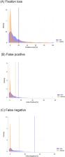
      Bookmark
          There is no author summary for this article yet. Authors can add summaries to their articles on ScienceOpen to make them more accessible to a non-specialist audience.

          Abstract

          This study compared between TEMPO, a new binocular perimeter, with the Humphrey Field Analyzer (HFA). Patients were tested with both TEMPO 24 − 2 AIZE-Rapid and HFA 24 − 2 SITA-Fast in a randomized sequence on the same day. Using a mixed-effects model, visual field (VF) parameters and reliability indices were compared. Retinal nerve fiber layer (RNFL) thickness was measured using Cirrus OCT, and coefficient of determinations for visual field and OCT parameters were calculated and compared using Akaike information criteria. 740 eyes (including 68 healthy, 262 glaucoma suspects, and 410 glaucoma) of 370 participants were evaluated. No significant differences were seen in mean deviation and visual field index between the two perimeters (P > 0.05). A stronger association between VF mean deviation and circumpapillary RNFL was found for TEMPO (adjusted R 2 = 0.28; AIC = 5210.9) compared to HFA (adjusted R 2 = 0.26; AIC = 5232.0). TEMPO had better reliability indices (fixation loss, false positive, and false negative) compared to HFA (all P < 0.05). Measurement time was faster for TEMPO compared to HFA (261sec vs. 429sec, P < 0.001). Further investigations are needed to assess the long-term monitoring potential of this binocular VF test.

          Related collections

          Most cited references27

          • Record: found
          • Abstract: found
          • Article: not found

          Primary open-angle glaucoma.

          Primary open-angle glaucoma is a progressive optic neuropathy and, perhaps, the most common form of glaucoma. Because the disease is treatable, and because the visual impairment caused by glaucoma is irreversible, early detection is essential. Early diagnosis depends on examination of the optic disc, retinal nerve fibre layer, and visual field. New imaging and psychophysical tests can improve both detection and monitoring of the progression of the disease. Recently completed long-term clinical trials provide convincing evidence that lowering intraocular pressure prevents progression at both the early and late stages of the disease. The degree of protection is related to the degree to which intraocular pressure is lowered. Improvements in therapy consist of more effective and better-tolerated drugs to lower intraocular pressure, and more effective surgical procedures. New treatments to directly treat and protect the retinal ganglion cells that are damaged in glaucoma are also in development.
            Bookmark
            • Record: found
            • Abstract: found
            • Article: not found

            Mapping the visual field to the optic disc in normal tension glaucoma eyes.

            To establish the anatomical relationship between visual field test points in the Humphrey 24-2 test pattern and regions of the optic nerve head (ONH) DESIGN: Cross-sectional study. Glaucoma patients and suspects from the Normal Tension Glaucoma Clinic at Moorfields Eye Hospital. Sixty-nine retinal nerve fiber layer (RNFL) photographs with well-defined RNFL defects and/or prominent bundles were digitized. An appropriately scaled Humphrey 24-2 visual field grid and an ONH reference circle, divided into 30 degrees sectors, were generated digitally. These were superimposed onto the RNFL images. The relationship of visual field test points to the circumference of the ONH was estimated by noting the proximity of test points to RNFL defects and/or prominent bundles. The position of the ONH in relation to the fovea was also noted. The sector at the ONH corresponding to each visual field test point, the position of the ONH in relation to the fovea, and the effect of the latter on the former. A median 22 (range, 4-58), of a possible 69, ONH positions were assigned to each visual field test point. The standard deviation of estimations was 7.2 degrees. The position of the ONH was 15.5 degrees (standard deviation 0.9 degrees ) nasal and 1.9 degrees (standard deviation 1.0 degrees ) above the fovea. The location of the ONH had a significant effect on the corresponding position at the ONH for 28 of 52 visual field test points. A clinically useful map that relates visual field test points to regions of the ONH has been produced. The map will aid clinical evaluation of glaucoma patients and suspects, as well as form the basis for investigations of the relationship between retinal light sensitivity and ONH structure.
              Bookmark
              • Record: found
              • Abstract: found
              • Article: not found

              Primary open-angle glaucoma.

              Glaucoma is an optic neuropathy that is characterized by the progressive degeneration of the optic nerve, leading to visual impairment. Glaucoma is the main cause of irreversible blindness worldwide, but typically remains asymptomatic until very severe. Open-angle glaucoma comprises the majority of cases in the United States and western Europe, of which, primary open-angle glaucoma (POAG) is the most common type. By contrast, in China and other Asian countries, angle-closure glaucoma is highly prevalent. These two types of glaucoma are characterized based on the anatomic configuration of the aqueous humour outflow pathway. The pathophysiology of POAG is not well understood, but it is an optic neuropathy that is thought to be associated with intraocular pressure (IOP)-related damage to the optic nerve head and resultant loss of retinal ganglion cells (RGCs). POAG is generally diagnosed during routine eye examination, which includes fundoscopic evaluation and visual field assessment (using perimetry). An increase in IOP, measured by tonometry, is not essential for diagnosis. Management of POAG includes topical drug therapies and surgery to reduce IOP, although new therapies targeting neuroprotection of RGCs and axonal regeneration are under development.
                Bookmark

                Author and article information

                Contributors
                Journal
                Res Sq
                ResearchSquare
                Research Square
                American Journal Experts
                29 August 2023
                : rs.3.rs-3283528
                Affiliations
                Hamilton Glaucoma Center, Shiley Eye Institute, University of California San Diego
                Hamilton Glaucoma Center, Shiley Eye Institute, University of California San Diego
                Hamilton Glaucoma Center, Shiley Eye Institute, University of California San Diego
                Hamilton Glaucoma Center, Shiley Eye Institute, University of California San Diego
                Hamilton Glaucoma Center, Shiley Eye Institute, University of California San Diego
                Author notes

                Author contributions

                Designed and conducted the study: T.N., R.N.W. and S.M. Data collection: T.N. and J.A., Data analysis: T.N. Interpretation: T.N., R.N.W. and S.M. Writing: T.N. and R.N.W. Critical revision: All authors. Manuscript approval: All authors.

                [] Correspondence and requests for materials should be addressed to S.M. sasanimii@ 123456yahoo.com
                Article
                10.21203/rs.3.rs-3283528
                10.21203/rs.3.rs-3283528/v1
                10491334
                37693597
                da81dab8-1583-400c-bc9a-7f87efb4e910

                This work is licensed under a Creative Commons Attribution 4.0 International License, which allows reusers to distribute, remix, adapt, and build upon the material in any medium or format, so long as attribution is given to the creator. The license allows for commercial use.

                History
                Funding
                Funded by: National Institutes of Health/National Eye Institute
                Award ID: R01EY029058
                Award ID: R01EY034148
                Categories
                Article

                Comments

                Comment on this article