43
views
0
recommends
+1 Recommend
0 collections
    0
    shares
      • Record: found
      • Abstract: found
      • Article: found
      Is Open Access

      Four Ca 2+ Ions Activate TRPM2 Channels by Binding in Deep Crevices near the Pore but Intracellularly of the Gate

      research-article
      ,
      The Journal of General Physiology
      The Rockefeller University Press

      Read this article at

      Bookmark
          There is no author summary for this article yet. Authors can add summaries to their articles on ScienceOpen to make them more accessible to a non-specialist audience.

          Abstract

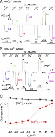
          TRPM2 is a tetrameric Ca 2+-permeable channel involved in immunocyte respiratory burst and in postischaemic neuronal death. In whole cells, TRPM2 activity requires intracellular ADP ribose (ADPR) and intra- or extracellular Ca 2+, but the mechanism and the binding sites for Ca 2+ activation remain unknown. Here we study TRPM2 gating in inside-out patches while directly controlling intracellular ligand concentrations. Concentration jump experiments at various voltages and Ca 2+ dependence of steady-state single-channel gating kinetics provide unprecedented insight into the molecular mechanism of Ca 2+ activation. In patches excised from Xenopus laevis oocytes expressing human TRPM2, coapplication of intracellular ADPR and Ca 2+ activated ∼50-pS nonselective cation channels; K 1/2 for ADPR was ∼1 µM at saturating Ca 2+. Intracellular Ca 2+ dependence of TRPM2 steady-state opening and closing rates (at saturating [ADPR] and low extracellular Ca 2+) reveals that Ca 2+ activation is a consequence of tighter binding of Ca 2+ in the open rather than in the closed channel conformation. Four Ca 2+ ions activate TRPM2 with a Monod-Wymann-Changeux mechanism: each binding event increases the open-closed equilibrium constant ∼33-fold, producing altogether 10 6-fold activation. Experiments in the presence of 1 mM of free Ca 2+ on the extracellular side clearly show that closed channels do not sense extracellular Ca 2+, but once channels have opened Ca 2+ entering passively through the pore slows channel closure by keeping the “activating sites” saturated, despite rapid continuous Ca 2+-free wash of the intracellular channel surface. This effect of extracellular Ca 2+ on gating is gradually lost at progressively depolarized membrane potentials, where the driving force for Ca 2+ influx is diminished. Thus, the activating sites lie intracellularly from the gate, but in a shielded crevice near the pore entrance. Our results suggest that in intact cells that contain micromolar ADPR a single brief puff of Ca 2+ likely triggers prolonged, self-sustained TRPM2 activity.

          Related collections

          Most cited references26

          • Record: found
          • Abstract: found
          • Article: not found

          Improved patch-clamp techniques for high-resolution current recording from cells and cell-free membrane patches.

          1. The extracellular patch clamp method, which first allowed the detection of single channel currents in biological membranes, has been further refined to enable higher current resolution, direct membrane patch potential control, and physical isolation of membrane patches. 2. A description of a convenient method for the fabrication of patch recording pipettes is given together with procedures followed to achieve giga-seals i.e. pipette-membrane seals with resistances of 10(9) - 10(11) omega. 3. The basic patch clamp recording circuit, and designs for improved frequency response are described along with the present limitations in recording the currents from single channels. 4. Procedures for preparation and recording from three representative cell types are given. Some properties of single acetylcholine-activated channels in muscle membrane are described to illustrate the improved current and time resolution achieved with giga-seals. 5. A description is given of the various ways that patches of membrane can be physically isolated from cells. This isolation enables the recording of single channel currents with well-defined solutions on both sides of the membrane. Two types of isolated cell-free patch configurations can be formed: an inside-out patch with its cytoplasmic membrane face exposed to the bath solution, and an outside-out patch with its extracellular membrane face exposed to the bath solution. 6. The application of the method for the recording of ionic currents and internal dialysis of small cells is considered. Single channel resolution can be achieved when recording from whole cells, if the cell diameter is small (less than 20 micrometer). 7. The wide range of cell types amenable to giga-seal formation is discussed.
            Bookmark
            • Record: found
            • Abstract: found
            • Article: not found

            Subunit stoichiometry of a mammalian K+ channel determined by construction of multimeric cDNAs.

            The subunit stoichiometry of the mammalian K+ channel KV1.1 (RCK1) was examined by linking together the coding sequences of 2-5 K+ channel subunits in a single open reading frame and tagging the expression of individual subunits with a mutation (Y379K or Y379R) that altered the sensitivity of the channel to block by external tetraethylammonium ion. Two lines of evidence argue that these constructs lead to K+ channel expression only through the formation of functional tetramers. First, currents expressed by tetrameric constructs containing a single mutant subunit have a sensitivity to tetraethylammonium that is well fitted by a single site binding isotherm. Second, a mutant subunit (Y379K) that expresses only as part of a heteromultimer contributes to the expression of functional channels when coexpressed with a trimeric construct but not a tetrameric construct.
              Bookmark
              • Record: found
              • Abstract: found
              • Article: not found

              ADP-ribose gating of the calcium-permeable LTRPC2 channel revealed by Nudix motif homology.

              Free ADP-ribose (ADPR), a product of NAD hydrolysis and a breakdown product of the calcium-release second messenger cyclic ADPR (cADPR), has no defined role as an intracellular signalling molecule in vertebrate systems. Here we show that a 350-amino-acid protein (designated NUDT9) and a homologous domain (NUDT9 homology domain) near the carboxy terminus of the LTRPC2/TrpC7 putative cation channel both function as specific ADPR pyrophosphatases. Whole-cell and single-channel analysis of HEK-293 cells expressing LTRPC2 show that LTRPC2 functions as a calcium-permeable cation channel that is specifically gated by free ADPR. The expression of native LTRPC2 transcripts is detectable in many tissues including the U937 monocyte cell line, in which ADPR induces large cation currents (designated IADPR) that closely match those mediated by recombinant LTRPC2. These results indicate that intracellular ADPR regulates calcium entry into cells that express LTRPC2.
                Bookmark

                Author and article information

                Journal
                J Gen Physiol
                J. Gen. Physiol
                jgp
                The Journal of General Physiology
                The Rockefeller University Press
                0022-1295
                1540-7748
                February 2009
                : 133
                : 2
                : 189-203
                Affiliations
                Department of Medical Biochemistry, Semmelweis University, Budapest, H-1094, Hungary
                Author notes
                Correspondence to László Csanády: laszlo.csanady@ 123456eok.sote.hu
                Article
                200810109
                10.1085/jgp.200810109
                2638199
                19171771
                dbc6ab20-eb41-4dfe-bb60-73c01d5b0064
                © 2009 Csanády and Törőcsik

                This article is distributed under the terms of an Attribution–Noncommercial–Share Alike–No Mirror Sites license for the first six months after the publication date (see http://www.jgp.org/misc/terms.shtml). After six months it is available under a Creative Commons License (Attribution–Noncommercial–Share Alike 3.0 Unported license, as described at http://creativecommons.org/licenses/by-nc-sa/3.0/).

                History
                : 21 August 2008
                : 6 January 2009
                Categories
                Article

                Anatomy & Physiology
                Anatomy & Physiology

                Comments

                Comment on this article