3
views
0
recommends
+1 Recommend
0 collections
    0
    shares
      • Record: found
      • Abstract: found
      • Article: found
      Is Open Access

      In Vitro Intraductal MRI and T2 Mapping of Cholangiocarcinoma Using Catheter Coils

      research-article

      Read this article at

      Bookmark
          There is no author summary for this article yet. Authors can add summaries to their articles on ScienceOpen to make them more accessible to a non-specialist audience.

          Abstract

          Aim

          Diagnostic imaging of early-stage cholangiocarcinoma is challenging. A previous in vitro study of fixed-tissue liver resection specimens investigated T2 mapping as a method of exploiting the locally increased signal-to-noise ratio (SNR) of duodenoscope coils for improved quantitative magnetic resonance imaging (MRI), despite their non-uniform sensitivity. This work applies similar methods to unfixed liver specimens using catheter-based receivers.

          Methods

          Ex vivo intraductal MRI and T2 mapping were carried out at 3T on unfixed resection specimens obtained from cholangiocarcinoma patients immediately after surgery using a catheter coil based on a thin-film magneto-inductive waveguide, inserted directly into an intrahepatic duct.

          Results

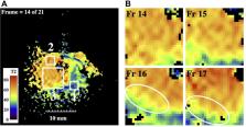
          Polypoid intraductal cholangiocarcinoma was imaged using fast spin-echo sequences. High-resolution T2 maps were extracted by fitting of data obtained at different echo times to mono-exponential models, and disease-induced changes were correlated with histopathology. An increase in T2 was found compared with fixed specimens and differences in T2 allowed the resolution of tumour tissue and malignant features such as polypoid morphology.

          Conclusion

          Despite their limited field of view, useful data can be obtained using catheter coils, and T2 mapping offers an effective method of exploiting their local SNR advantage without the need for image correction.

          Related collections

          Most cited references34

          • Record: found
          • Abstract: found
          • Article: not found

          Cholangiocarcinoma.

          Cholangiocarcinoma is a devastating malignancy that presents late, is notoriously difficult to diagnose, and is associated with a high mortality. The incidence of intrahepatic cholangiocarcinoma is increasing worldwide. The cause for this rise is unclear, although it could be related to an interplay between predisposing genetic factors and environmental triggers. MRI and CT with endoscopic ultrasound and PET provide useful diagnostic information in certain patients. Surgical resection is the only chance for cure, with results depending on careful technique and patient selection. Data suggest that liver transplantation could offer long-term survival in selected patients when combined with neoadjuvant chemoradiotherapy. Chemotherapy and radiotherapy have been ineffective for patients with inoperable tumours. For most of these patients biliary drainage is the mainstay of palliation. However, controversy exists over the type and positioning of biliary stents. Photodynamic treatment is a new palliative technique that might improve quality of life.
            Bookmark
            • Record: found
            • Abstract: found
            • Article: not found

            The tumorigenic liver fluke Opisthorchis viverrini--multiple pathways to cancer.

            Liver fluke infection caused by Opisthorchis viverrini is a major public health problem in Thailand and adjacent countries. In addition to infection-associated morbidity, infection with O. viverrini and the related Clonorchis sinensis are unarguable risk factors for cholangiocarcinoma (CAA, bile-duct cancer). Here we review the pathogenesis of opisthorchiasis and the association between O. viverrini infection and bile-duct cancer, focusing on the molecular parallels between wound healing, chronic inflammation, and cancer development. We review a schema for human disease progression from fluke infection, chronic opisthorchiasis, advanced periductal fibrosis, and cholangiocarcinogenesis, and present a rationale for biomarker discovery to facilitate early intervention. We conclude by addressing post-genomic advances with a view to developing new control strategies to combat this infectious cancer. Copyright © 2012 Elsevier Ltd. All rights reserved.
              Bookmark
              • Record: found
              • Abstract: found
              • Article: not found

              Histological diversity in cholangiocellular carcinoma reflects the different cholangiocyte phenotypes.

              Cholangiocellular carcinoma (CC) originates from topographically heterogeneous cholangiocytes. The cylindrical mucin-producing cholangiocytes are located in large bile ducts and the cuboidal non-mucin-producing cholangiocytes are located in ductules containing bipotential hepatic progenitor cells (HPCs). We investigated the clinicopathological and molecular features of 85 resected CCs (14 hilar CCs [so-called Klatskin tumor], 71 intrahepatic CCs [ICCs] including 20 cholangiolocellular carcinomas [CLCs], which are thought to originate from HPCs]) and compared these with the different cholangiocyte phenotypes, including HPCs. Immunohistochemistry was performed with biliary/HPC and hepatocytic markers. Gene expression profiling was performed in different tumors and compared with nonneoplastic different cholangiocyte phenotypes obtained by laser microdissection. Invasion and cell proliferation assay were assessed using different types of CC cell lines: KMC-1, KMCH-1, and KMCH-2. Among 51 ICCs, 31 (60.8%) contained only mucin-producing CC features (muc-ICCs), whereas 39.2% displayed histological diversity: focal hepatocytic differentiation and ductular areas (mixed-ICCs). Clinicopathologically, muc-ICCs and hilar CCs showed a predominantly (peri-)hilar location, smaller tumor size, and more lymphatic and perineural invasion compared with mixed-ICCs and CLCs (predominantly peripheral location, larger tumor size, and less lymphatic and perineural invasion). Immunoreactivity was similar in muc-ICCs and hilar CCs and in mixed-ICCs and CLCs. S100P and MUC1 were significantly up-regulated in hilar CCs and muc-ICCs compared with mixed-ICCs and CLCs, whereas NCAM1 and ALB tended to be up-regulated in mixed-ICCs and CLCs compared with other tumors. KMC-1 showed significantly higher invasiveness than KMCH-1 and KMCH-2. Muc-ICCs had a clinicopathological, immunohistochemical, and molecular profile similar to that of hilar CCs (from mucin-producing cholangiocytes), whereas mixed-ICCs had a profile similar to that of CLCs (thought to be of HPC origin), possibly reflecting their respective cells of origin. Copyright © 2012 American Association for the Study of Liver Diseases.
                Bookmark

                Author and article information

                Journal
                Hepat Med
                Hepat Med
                HMER
                hmer
                Hepatic Medicine : Evidence and Research
                Dove
                1179-1535
                27 July 2020
                2020
                : 12
                : 107-114
                Affiliations
                [1 ]Department. of Surgery, Faculty of Medicine, Khon Kaen University , Khon Kaen, 40002, Thailand
                [2 ]Department of Radiology, Faculty of Medicine, Khon Kaen University , Khon Kaen 40002, Thailand
                [3 ]Department of Pathology, Faculty of Medicine, Khon Kaen University , Khon Kaen 40002, Thailand
                [4 ]Division of Surgery and Cancer, Imperial College London , Paddington, London W2 1NY, UK
                [5 ]Philips Healthcare Germany, Health Systems, Clinical Science , Hamburg, Germany
                [6 ]EEE Department, Imperial College , London SW7 2AZ, UK
                Author notes
                Correspondence: Simon D Taylor-Robinson Division of Surgery and Cancer, Imperial College London , Liver Unit, St. Mary’s Hospital, South Wharf Road, Paddington, LondonW2 1NY, UKTel +44 20 3312 6199 Email s.taylor-robinson@imperial.ac.uk
                Author information
                http://orcid.org/0000-0001-9305-0688
                http://orcid.org/0000-0002-4117-2330
                http://orcid.org/0000-0002-1057-7255
                http://orcid.org/0000-0003-0901-8314
                Article
                266841
                10.2147/HMER.S266841
                7397475
                dbd4bcf0-41f8-4288-a183-d56256c1ce08
                © 2020 Khuntikeo et al.

                This work is published by Dove Medical Press Limited, and licensed under a Creative Commons Attribution License. The full terms of the License are available at http://creativecommons.org/licenses/by/4.0/. The license permits unrestricted use, distribution, and reproduction in any medium, provided the original author and source are credited.

                History
                : 09 June 2020
                : 09 July 2020
                Page count
                Figures: 5, References: 40, Pages: 8
                Categories
                Original Research

                cholangiocarcinoma,t2 mapping,catheter coil
                cholangiocarcinoma, t2 mapping, catheter coil

                Comments

                Comment on this article