0
views
0
recommends
+1 Recommend
0 collections
    0
    shares
      • Record: found
      • Abstract: found
      • Article: found
      Is Open Access

      A multicenter study of modified electron beam output calibration

      research-article

      Read this article at

      Bookmark
          There is no author summary for this article yet. Authors can add summaries to their articles on ScienceOpen to make them more accessible to a non-specialist audience.

          Abstract

          Purpose

          This study aims to assess the accuracy of a modified electron beam calibration based on the IAEA TRS‐398 and AAPM‐TG‐51 in multicenter radiotherapy.

          Methods

          This study was performed using the Elekta and Varian Linear Accelerator electron beams with energies of 4–22 MeV under reference conditions using cylindrical (PTW 30013, IBA FC65‐G, and IBA FC65‐P) and parallel‐plate (PTW 34045, PTW 34001, and IBA PPC‐40) chambers. The modified calibration used a cylindrical chamber and an updated k Q based on Monte Carlo calculations, whereas TRS‐398 and TG‐51 used cylindrical and parallel‐plate chambers for reference dosimetry. The dose ratio of the modified calibration procedure, TRS‐398 and TG‐51 were obtained by comparing the dose at the maximum depth of the modified calibration to TRS‐398 and TG‐51.

          Results

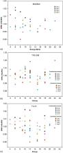
          The study found that all cylindrical chambers’ beam quality conversion factors determined with the modified calibration ( k Q ) to the TRS‐398 and TG‐51 vary from 0.994 to 1.003 and 1.000 to 1.010, respectively. The dose ratio of modified/TRS‐398 cyl and modified/TRS‐398 parallel‐plate, the variation ranges were 0.980–1.014 and 0.981–1.019, while for the counterpart modified/TG‐51 cyl was found varying between 0.991 and 1.017 and the ratio of modified/TG‐51 parallel‐plate varied in the range of 0.981–1.019.

          Conclusion

          This multi‐institutional study analyzed a modified calibration procedure utilizing new data for electron beam calibrations at multiple institutions and evaluated existing calibration protocols. Based on observed variations, the current calibration protocols should be updated with detailed metrics on the stability of linac components.

          Related collections

          Most cited references25

          • Record: found
          • Abstract: found
          • Article: not found

          AAPM's TG-51 protocol for clinical reference dosimetry of high-energy photon and electron beams.

          A protocol is prescribed for clinical reference dosimetry of external beam radiation therapy using photon beams with nominal energies between 60Co and 50 MV and electron beams with nominal energies between 4 and 50 MeV. The protocol was written by Task Group 51 (TG-51) of the Radiation Therapy Committee of the American Association of Physicists in Medicine (AAPM) and has been formally approved by the AAPM for clinical use. The protocol uses ion chambers with absorbed-dose-to-water calibration factors, N(60Co)D,w which are traceable to national primary standards, and the equation D(Q)w = MkQN(60Co)D,w where Q is the beam quality of the clinical beam, D(Q)w is the absorbed dose to water at the point of measurement of the ion chamber placed under reference conditions, M is the fully corrected ion chamber reading, and kQ is the quality conversion factor which converts the calibration factor for a 60Co beam to that for a beam of quality Q. Values of kQ are presented as a function of Q for many ion chambers. The value of M is given by M = PionP(TP)PelecPpolMraw, where Mraw is the raw, uncorrected ion chamber reading and Pion corrects for ion recombination, P(TP) for temperature and pressure variations, Pelec for inaccuracy of the electrometer if calibrated separately, and Ppol for chamber polarity effects. Beam quality, Q, is specified (i) for photon beams, by %dd(10)x, the photon component of the percentage depth dose at 10 cm depth for a field size of 10x10 cm2 on the surface of a phantom at an SSD of 100 cm and (ii) for electron beams, by R50, the depth at which the absorbed-dose falls to 50% of the maximum dose in a beam with field size > or =10x10 cm2 on the surface of the phantom (> or =20x20 cm2 for R50>8.5 cm) at an SSD of 100 cm. R50 is determined directly from the measured value of I50, the depth at which the ionization falls to 50% of its maximum value. All clinical reference dosimetry is performed in a water phantom. The reference depth for calibration purposes is 10 cm for photon beams and 0.6R50-0.1 cm for electron beams. For photon beams clinical reference dosimetry is performed in either an SSD or SAD setup with a 10x10 cm2 field size defined on the phantom surface for an SSD setup or at the depth of the detector for an SAD setup. For electron beams clinical reference dosimetry is performed with a field size of > or =10x10 cm2 (> or =20x20 cm2 for R50>8.5 cm) at an SSD between 90 and 110 cm. This protocol represents a major simplification compared to the AAPM's TG-21 protocol in the sense that large tables of stopping-power ratios and mass-energy absorption coefficients are not needed and the user does not need to calculate any theoretical dosimetry factors. Worksheets for various situations are presented along with a list of equipment required.
            Bookmark
            • Record: found
            • Abstract: not found
            • Book: not found

            Introduction to Radiological Physics and Radiation Dosimetry

              Bookmark
              • Record: found
              • Abstract: found
              • Article: not found

              Addendum to the AAPMˈs TG-51 protocol for clinical reference dosimetry of high-energy photon beams

              An addendum to the AAPM's TG-51 protocol for the determination of absorbed dose to water in megavoltage photon beams is presented. This addendum continues the procedure laid out in TG-51 but new k Q data for photon beams, based on Monte Carlo simulations, are presented and recommendations are given to improve the accuracy and consistency of the protocol's implementation. The components of the uncertainty budget in determining absorbed dose to water at the reference point are introduced and the magnitude of each component discussed. Finally, the consistency of experimental determination of N D,w coefficients is discussed. It is expected that the implementation of this addendum will be straightforward, assuming that the user is already familiar with TG-51. The changes introduced by this report are generally minor, although new recommendations could result in procedural changes for individual users. It is expected that the effort on the medical physicist's part to implement this addendum will not be significant and could be done as part of the annual linac calibration.
                Bookmark

                Author and article information

                Contributors
                supriyanto.p@sci.ui.ac.id
                Journal
                J Appl Clin Med Phys
                J Appl Clin Med Phys
                10.1002/(ISSN)1526-9914
                ACM2
                Journal of Applied Clinical Medical Physics
                John Wiley and Sons Inc. (Hoboken )
                1526-9914
                13 December 2023
                January 2024
                : 25
                : 1 ( doiID: 10.1002/acm2.v25.1 )
                : e14232
                Affiliations
                [ 1 ] Department of Physics Faculty of Mathematics and Natural Sciences Universitas Indonesia, Depok West Java Indonesia
                [ 2 ] Department of Radiation Oncology Mitra Keluarga Bekasi Timur Hospital, Bekasi West Java Indonesia
                [ 3 ] Department of Radiation Oncology Gading Pluit Hospital Jakarta Indonesia
                [ 4 ] Department of Radiation Oncology Persahabatan Central General Hospital Jakarta Indonesia
                [ 5 ] Department of Radiation Oncology Mayapada Hospital Jakarta Selatan Jakarta Indonesia
                [ 6 ] Department of Radiation Oncology MRCCC Siloam Hospital Semanggi Jakarta Indonesia
                [ 7 ] Department of Radiation Oncology Pasar Minggu Regional Hospital Jakarta Indonesia
                [ 8 ] Department of Radiation Oncology Siloam Hospital TB Simatupang Jakarta Indonesia
                [ 9 ] Department of Radiation Oncology Tzu Chi Hospital Jakarta Indonesia
                [ 10 ] Department of Radiation Oncology Dr. Cipto Mangunkusumo National General Hospital Central Faculty of Medicine Universitas Indonesia Jakarta Indonesia
                [ 11 ] Department of Radiation Oncology Northwestern Memorial Hospital Northwestern University Feinberg School of Medicine Chicago Illinois USA
                Author notes
                [*] [* ] Correspondence

                Supriyanto Ardjo Pawiro, Department of Physics, Faculty of Mathematics and Natural Sciences, Universitas Indonesia, Depok, West Java, Indonesia.

                Email: supriyanto.p@ 123456sci.ui.ac.id

                Author information
                https://orcid.org/0000-0002-0028-1442
                Article
                ACM214232
                10.1002/acm2.14232
                10795448
                38088260
                dbeb7ec0-00ff-4c4e-88d4-a72a67441d94
                © 2023 The Authors. Journal of Applied Clinical Medical Physics published by Wiley Periodicals LLC on behalf of American Association of Physicists in Medicine.

                This is an open access article under the terms of the http://creativecommons.org/licenses/by/4.0/ License, which permits use, distribution and reproduction in any medium, provided the original work is properly cited.

                History
                : 13 November 2023
                : 28 February 2023
                : 19 November 2023
                Page count
                Figures: 3, Tables: 7, Pages: 10, Words: 5629
                Categories
                Radiation Measurements
                Radiation Measurements
                Custom metadata
                2.0
                January 2024
                Converter:WILEY_ML3GV2_TO_JATSPMC version:6.3.6 mode:remove_FC converted:18.01.2024

                beam quality conversion factor,electron reference dosimetry,modified calibration,monte carlo simulation,trs‐398 protocol

                Comments

                Comment on this article