13
views
0
recommends
+1 Recommend
0 collections
    0
    shares
      • Record: found
      • Abstract: found
      • Article: not found

      Plate Tectonics of Virus Shell Assembly and Reorganization in Phage Φ8, a Distant Relative of Mammalian Reoviruses

      research-article

      Read this article at

      ScienceOpenPublisherPMC
      Bookmark
          There is no author summary for this article yet. Authors can add summaries to their articles on ScienceOpen to make them more accessible to a non-specialist audience.

          Summary

          The hallmark of a virus is its capsid, which harbors the viral genome and is formed from protein subunits, which assemble following precise geometric rules. dsRNA viruses use an unusual protein multiplicity (120 copies) to form their closed capsids. We have determined the atomic structure of the capsid protein (P1) from the dsRNA cystovirus Φ8. In the crystal P1 forms pentamers, very similar in shape to facets of empty procapsids, suggesting an unexpected assembly pathway that proceeds via a pentameric intermediate. Unlike the elongated proteins used by dsRNA mammalian reoviruses, P1 has a compact trapezoid-like shape and a distinct arrangement in the shell, with two near-identical conformers in nonequivalent structural environments. Nevertheless, structural similarity with the analogous protein from the mammalian viruses suggests a common ancestor. The unusual shape of the molecule may facilitate dramatic capsid expansion during phage maturation, allowing P1 to switch interaction interfaces to provide capsid plasticity.
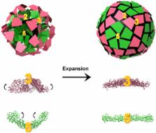
          Graphical Abstract

          Highlights

          • Crystal structure of the major capsid protein P1 of the Pseudomonas phage Φ8

          • Φ8 P1 shares a common ancestor with mammalian reoviruses

          • Φ8 P1’s trapezoidal shape may facilitate capsid expansion during maturation

          • The pentameric organization of Φ8 P1 suggests a revised assembly pathway

          Abstract

          El Omari et al. report a structure of the dsRNA bacteriophage ϕ8 capsid protein P1. P1 crystallizes as a pentamer, suggesting a new pathway for procapsid assembly. P1 displays a novel fold and a trapezoidal shape, distinct from that of other dsRNA virus, which may facilitate capsid expansion during maturation.

          Related collections

          Most cited references55

          • Record: found
          • Abstract: found
          • Article: not found

          Structure unifies the viral universe.

          Is it possible to meaningfully comprehend the diversity of the viral world? We propose that it is. This is based on the observation that, although there is immense genomic variation, every infective virion is restricted by strict constraints in structure space (i.e., there are a limited number of ways to fold a protein chain, and only a small subset of these have the potential to construct a virion, the hallmark of a virus). We have previously suggested the use of structure for the higher-order classification of viruses, where genomic similarities are no longer observable. Here, we summarize the arguments behind this proposal, describe the current status of structural work, highlighting its power to infer common ancestry, and discuss the limitations and obstacles ahead of us. We also reflect on the future opportunities for a more concerted effort to provide high-throughput methods to facilitate the large-scale sampling of the virosphere.
            Bookmark
            • Record: found
            • Abstract: found
            • Article: not found

            A mechanism for initiating RNA-dependent RNA polymerization.

            In most RNA viruses, genome replication and transcription are catalysed by a viral RNA-dependent RNA polymerase. Double-stranded RNA viruses perform these operations in a capsid (the polymerase complex), using an enzyme that can read both single- and double-stranded RNA. Structures have been solved for such viral capsids, but they do not resolve the polymerase subunits in any detail. Here we show that the 2 A resolution X-ray structure of the active polymerase subunit from the double-stranded RNA bacteriophage straight phi6 is highly similar to that of the polymerase of hepatitis C virus, providing an evolutionary link between double-stranded RNA viruses and flaviviruses. By crystal soaking and co-crystallization, we determined a number of other structures, including complexes with oligonucleotide and/or nucleoside triphosphates (NTPs), that suggest a mechanism by which the incoming double-stranded RNA is opened up to feed the template through to the active site, while the substrates enter by another route. The template strand initially overshoots, locking into a specificity pocket, and then, in the presence of cognate NTPs, reverses to form the initiation complex; this process engages two NTPs, one of which acts with the carboxy-terminal domain of the protein to prime the reaction. Our results provide a working model for the initiation of replication and transcription.
              Bookmark
              • Record: found
              • Abstract: not found
              • Article: not found

              Crystal structure of cat muscle pyruvate kinase at a resolution of 2.6 A.

                Bookmark

                Author and article information

                Contributors
                Journal
                Structure
                Structure
                Structure(London, England:1993)
                Cell Press
                0969-2126
                1878-4186
                06 August 2013
                06 August 2013
                : 21
                : 8
                : 1384-1395
                Affiliations
                [1 ]Division of Structural Biology, The Wellcome Trust Centre for Human Genetics, University of Oxford, Headington, Oxford OX3 7BN, UK
                [2 ]Institute of Biotechnology and Department of Biosciences, University of Helsinki, Biocenter 2, P.O. Box 56, 00014 Helsinki, Finland
                [3 ]Diamond Light Source Limited, Harwell Science and Innovation Campus, Didcot OX11 0DE, UK
                Author notes
                []Corresponding author erika@ 123456strubi.ox.ac.uk
                Article
                STFODE2733
                10.1016/j.str.2013.06.017
                3737474
                23891291
                dbf6ae7f-d061-4f55-8626-ab48024ff7f7
                © 2013 The Authors

                This document may be redistributed and reused, subject to certain conditions.

                History
                : 11 May 2013
                : 26 June 2013
                : 27 June 2013
                Categories
                Article

                Molecular biology
                Molecular biology

                Comments

                Comment on this article