0
views
0
recommends
+1 Recommend
0 collections
    0
    shares
      • Record: found
      • Abstract: found
      • Article: found
      Is Open Access

      Functional and structural alterations of dorsal attention network in preclinical and early‐stage Alzheimer's disease

      research-article

      Read this article at

      Bookmark
          There is no author summary for this article yet. Authors can add summaries to their articles on ScienceOpen to make them more accessible to a non-specialist audience.

          Abstract

          Objectives

          Subjective cognitive decline (SCD) and amnestic mild cognitive impairment (aMCI) are known as the preclinical and early stage of Alzheimer's disease (AD). The dorsal attention network (DAN) is mainly responsible for the “top‐down” attention process. However, previous studies mainly focused on single functional modality and limited structure. This study aimed to investigate the multimodal alterations of DAN in SCD and aMCI to assess their diagnostic value in preclinical and early‐stage AD.

          Methods

          Resting‐state functional magnetic resonance imaging (MRI) was carried out to measure the fractional amplitude of low‐frequency fluctuation (fALFF), regional homogeneity (ReHo), and functional connectivity (FC). Structural MRI was used to calculate the gray matter volume (GMV) and cortical thickness. Moreover, receiver‐operating characteristic (ROC) analysis was used to distinguish these alterations in SCD and aMCI.

          Results

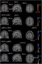
          The SCD and aMCI groups showed both decreased ReHo in the right middle temporal gyrus (MTG) and decreased GMV compared to healthy controls (HCs). Especially in the SCD group, there were increased fALFF and increased ReHo in the left inferior occipital gyrus (IOG), decreased fALFF and increased FC in the left inferior parietal lobule (IPL), and reduced cortical thickness in the right inferior temporal gyrus (ITG). Furthermore, functional and structural alterations in the SCD and aMCI groups were closely related to episodic memory (EM), executive function (EF), and information processing speed (IPS). The combination of multiple indicators of DAN had a high accuracy in differentiating clinical stages.

          Conclusions

          Our current study demonstrated functional and structural alterations of DAN in SCD and aMCI, especially in the MTG, IPL, and SPL. Furthermore, cognitive performance was closely related to these significant alterations. Our study further suggested that the combined multiple indicators of DAN could be acted as the latent neuroimaging markers of preclinical and early‐stage AD for their high diagnostic value.

          Abstract

          Our current study demonstrated obviously functional and structural alterations of DAN in SCD and aMCI. We found that the abnormalities of the functional alterations were especially located in the IPL, IOG, and MTG, whereas structural alterations were mainly in the SPL and ITG. Furthermore, cognitive performance was closely related to these significant alterations. Our study further suggested that the combined multiple indicators of DAN could be acted as the latent neuroimaging markers of preclinical and early‐stage AD for their high diagnostic value.

          Related collections

          Most cited references66

          • Record: found
          • Abstract: found
          • Article: not found

          DPABI: Data Processing & Analysis for (Resting-State) Brain Imaging.

          Brain imaging efforts are being increasingly devoted to decode the functioning of the human brain. Among neuroimaging techniques, resting-state fMRI (R-fMRI) is currently expanding exponentially. Beyond the general neuroimaging analysis packages (e.g., SPM, AFNI and FSL), REST and DPARSF were developed to meet the increasing need of user-friendly toolboxes for R-fMRI data processing. To address recently identified methodological challenges of R-fMRI, we introduce the newly developed toolbox, DPABI, which was evolved from REST and DPARSF. DPABI incorporates recent research advances on head motion control and measurement standardization, thus allowing users to evaluate results using stringent control strategies. DPABI also emphasizes test-retest reliability and quality control of data processing. Furthermore, DPABI provides a user-friendly pipeline analysis toolkit for rat/monkey R-fMRI data analysis to reflect the rapid advances in animal imaging. In addition, DPABI includes preprocessing modules for task-based fMRI, voxel-based morphometry analysis, statistical analysis and results viewing. DPABI is designed to make data analysis require fewer manual operations, be less time-consuming, have a lower skill requirement, a smaller risk of inadvertent mistakes, and be more comparable across studies. We anticipate this open-source toolbox will assist novices and expert users alike and continue to support advancing R-fMRI methodology and its application to clinical translational studies.
            Bookmark
            • Record: found
            • Abstract: found
            • Article: not found

            A conceptual framework for research on subjective cognitive decline in preclinical Alzheimer's disease

            There is increasing evidence that subjective cognitive decline (SCD) in individuals with unimpaired performance on cognitive tests may represent the first symptomatic manifestation of Alzheimer's disease (AD). The research on SCD in early AD, however, is limited by the absence of common standards. The working group of the Subjective Cognitive Decline Initiative (SCD-I) addressed this deficiency by reaching consensus on terminology and on a conceptual framework for research on SCD in AD. In this publication, research criteria for SCD in pre-mild cognitive impairment (MCI) are presented. In addition, a list of core features proposed for reporting in SCD studies is provided, which will enable comparability of research across different settings. Finally, a set of features is presented, which in accordance with current knowledge, increases the likelihood of the presence of preclinical AD in individuals with SCD. This list is referred to as SCD plus.
              Bookmark
              • Record: found
              • Abstract: found
              • Article: found
              Is Open Access

              DPARSF: A MATLAB Toolbox for “Pipeline” Data Analysis of Resting-State fMRI

              Resting-state functional magnetic resonance imaging (fMRI) has attracted more and more attention because of its effectiveness, simplicity and non-invasiveness in exploration of the intrinsic functional architecture of the human brain. However, user-friendly toolbox for “pipeline” data analysis of resting-state fMRI is still lacking. Based on some functions in Statistical Parametric Mapping (SPM) and Resting-State fMRI Data Analysis Toolkit (REST), we have developed a MATLAB toolbox called Data Processing Assistant for Resting-State fMRI (DPARSF) for “pipeline” data analysis of resting-state fMRI. After the user arranges the Digital Imaging and Communications in Medicine (DICOM) files and click a few buttons to set parameters, DPARSF will then give all the preprocessed (slice timing, realign, normalize, smooth) data and results for functional connectivity, regional homogeneity, amplitude of low-frequency fluctuation (ALFF), and fractional ALFF. DPARSF can also create a report for excluding subjects with excessive head motion and generate a set of pictures for easily checking the effect of normalization. In addition, users can also use DPARSF to extract time courses from regions of interest.
                Bookmark

                Author and article information

                Contributors
                linxingjian@njmu.edu.cn
                ericcst@aliyun.com
                Journal
                CNS Neurosci Ther
                CNS Neurosci Ther
                10.1111/(ISSN)1755-5949
                CNS
                CNS Neuroscience & Therapeutics
                John Wiley and Sons Inc. (Hoboken )
                1755-5930
                1755-5949
                21 March 2023
                June 2023
                : 29
                : 6 ( doiID: 10.1002/cns.v29.6 )
                : 1512-1524
                Affiliations
                [ 1 ] Department of Neurology The Affiliated Brain Hospital of Nanjing Medical University Nanjing China
                [ 2 ] Institute of Neuropsychiatry The Affiliated Brain Hospital of Nanjing Medical University Nanjing China
                [ 3 ] Institute of Brain Functional Imaging Nanjing Medical University Nanjing China
                [ 4 ] Department of Radiology The Affiliated Brain Hospital of Nanjing Medical University Nanjing China
                [ 5 ] Department of Radiology, Affiliated Drum Tower Hospital Medical School of Nanjing University Nanjing China
                [ 6 ] Institute of Medical Imaging and Artificial Intelligence Nanjing University Nanjing China
                [ 7 ] Medical Imaging Center, Affiliated Drum Tower Hospital Medical School of Nanjing University Nanjing China
                Author notes
                [*] [* ] Correspondence

                Xingjian Lin, Department of Neurology, The Affiliated Brain Hospital of Nanjing Medical University, No. 264, Guangzhou Road, Gulou District, Nanjing, Jiangsu 210029, China.

                Email: linxingjian@ 123456njmu.edu.cn

                Jiu Chen, Department of Radiology, Affiliated Drum Tower Hospital, Medical School of Nanjing University, No. 281, Zhongshan Road, Gulou District, Nanjing, Jiangsu 210029, China.

                Email: ericcst@ 123456aliyun.com

                Author information
                https://orcid.org/0000-0002-2675-5837
                https://orcid.org/0000-0001-8185-8575
                Article
                CNS14092 CNSNT-2022-748.R2
                10.1111/cns.14092
                10173716
                36942514
                deffb9d5-3443-4d59-847d-45c45cf05296
                © 2023 The Authors. CNS Neuroscience & Therapeutics published by John Wiley & Sons Ltd.

                This is an open access article under the terms of the http://creativecommons.org/licenses/by/4.0/ License, which permits use, distribution and reproduction in any medium, provided the original work is properly cited.

                History
                : 31 December 2022
                : 18 September 2022
                : 02 January 2023
                Page count
                Figures: 7, Tables: 6, Pages: 13, Words: 7387
                Funding
                Funded by: Key Research and Development Program of Jiangxi Province , doi 10.13039/501100013064;
                Award ID: BE2022679
                Award ID: BE2018608
                Funded by: Major Basic Research Project of the Natural Science Foundation of the Jiangsu Higher Education Institutions , doi 10.13039/501100013280;
                Award ID: BK20221185
                Funded by: National Natural Science Foundation of China , doi 10.13039/501100001809;
                Award ID: 81701675
                Funded by: Special Funded Project of Nanjing Drum Tower Hospital
                Award ID: RC2022‐023
                Categories
                Original Article
                Original Articles
                Custom metadata
                2.0
                June 2023
                Converter:WILEY_ML3GV2_TO_JATSPMC version:6.2.8 mode:remove_FC converted:11.05.2023

                Neurosciences
                amnestic mild cognitive impairment,dorsal attention network,resting‐state functional magnetic resonance imaging,structural magnetic resonance imaging,subjective cognitive decline

                Comments

                Comment on this article