1
views
0
recommends
+1 Recommend
0 collections
    0
    shares
      • Record: found
      • Abstract: found
      • Article: found
      Is Open Access

      Unified Topological Inference for Brain Networks in Temporal Lobe Epilepsy Using the Wasserstein Distance

      Preprint
      research-article

      Read this article at

      Bookmark
          There is no author summary for this article yet. Authors can add summaries to their articles on ScienceOpen to make them more accessible to a non-specialist audience.

          Abstract

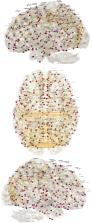
          Persistent homology offers a powerful tool for extracting hidden topological signals from brain networks. It captures the evolution of topological structures across multiple scales, known as filtrations, thereby revealing topological features that persist over these scales. These features are summarized in persistence diagrams, and their dissimilarity is quantified using the Wasserstein distance. However, the Wasserstein distance does not follow a known distribution, posing challenges for the application of existing parametric statistical models. To tackle this issue, we introduce a unified topological inference framework centered on the Wasserstein distance. Our approach has no explicit model and distributional assumptions. The inference is performed in a completely data driven fashion. We apply this method to resting-state functional magnetic resonance images (rs-fMRI) of temporal lobe epilepsy patients collected from two different sites: the University of Wisconsin-Madison and the Medical College of Wisconsin. Importantly, our topological method is robust to variations due to sex and image acquisition, obviating the need to account for these variables as nuisance covariates. We successfully localize the brain regions that contribute the most to topological differences. A MATLAB package used for all analyses in this study is available at https://github.com/laplcebeltrami/PH-STAT.

          Related collections

          Most cited references116

          • Record: found
          • Abstract: found
          • Article: not found

          The minimal preprocessing pipelines for the Human Connectome Project.

          The Human Connectome Project (HCP) faces the challenging task of bringing multiple magnetic resonance imaging (MRI) modalities together in a common automated preprocessing framework across a large cohort of subjects. The MRI data acquired by the HCP differ in many ways from data acquired on conventional 3 Tesla scanners and often require newly developed preprocessing methods. We describe the minimal preprocessing pipelines for structural, functional, and diffusion MRI that were developed by the HCP to accomplish many low level tasks, including spatial artifact/distortion removal, surface generation, cross-modal registration, and alignment to standard space. These pipelines are specially designed to capitalize on the high quality data offered by the HCP. The final standard space makes use of a recently introduced CIFTI file format and the associated grayordinate spatial coordinate system. This allows for combined cortical surface and subcortical volume analyses while reducing the storage and processing requirements for high spatial and temporal resolution data. Here, we provide the minimum image acquisition requirements for the HCP minimal preprocessing pipelines and additional advice for investigators interested in replicating the HCP's acquisition protocols or using these pipelines. Finally, we discuss some potential future improvements to the pipelines. Copyright © 2013 Elsevier Inc. All rights reserved.
            Bookmark
            • Record: found
            • Abstract: found
            • Article: not found

            Whole brain segmentation: automated labeling of neuroanatomical structures in the human brain.

            We present a technique for automatically assigning a neuroanatomical label to each voxel in an MRI volume based on probabilistic information automatically estimated from a manually labeled training set. In contrast to existing segmentation procedures that only label a small number of tissue classes, the current method assigns one of 37 labels to each voxel, including left and right caudate, putamen, pallidum, thalamus, lateral ventricles, hippocampus, and amygdala. The classification technique employs a registration procedure that is robust to anatomical variability, including the ventricular enlargement typically associated with neurological diseases and aging. The technique is shown to be comparable in accuracy to manual labeling, and of sufficient sensitivity to robustly detect changes in the volume of noncortical structures that presage the onset of probable Alzheimer's disease.
              Bookmark
              • Record: found
              • Abstract: found
              • Article: not found

              Efficient Behavior of Small-World Networks

              We introduce the concept of efficiency of a network as a measure of how efficiently it exchanges information. By using this simple measure, small-world networks are seen as systems that are both globally and locally efficient. This gives a clear physical meaning to the concept of "small world," and also a precise quantitative analysis of both weighted and unweighted networks. We study neural networks and man-made communication and transportation systems and we show that the underlying general principle of their construction is in fact a small-world principle of high efficiency.
                Bookmark

                Author and article information

                Contributors
                Journal
                ArXiv
                ArXiv
                arxiv
                ArXiv
                Cornell University
                2331-8422
                20 September 2023
                : arXiv:2302.06673v3
                Affiliations
                Department of Biostatistics and Medical Informatics, University of Wisconsin-Madison, USA
                Department of Neurology, University of Wisconsin-Madison, USA
                Department of Neurology, University of Wisconsin-Madison, USA
                Department of Neurology, Medical College of Wisconsin, USA
                Department of Radiology, University of Wisconsin-Madison, USA
                Department of Radiology, University of Wisconsin-Madison, USA
                Departments of Medical Physics & Biomedical Engineering, University of Wisconsin-Madison, USA
                Department of Neurology, University of Wisconsin-Madison, USA
                Department of Neurology, Medical College of Wisconsin, USA
                Department of Neurology, University of Wisconsin-Madison, USA
                Author notes
                Moo K. Chung, Department of Biostatistics and Medical Informatics, University of Wisconsin-Madison, USA mkchung@ 123456wisc.edu
                Article
                2302.06673
                9949148
                36824424
                df1c5036-baf1-4df5-8ada-b53fd4cf9481

                This work is licensed under a Creative Commons Attribution-NonCommercial-ShareAlike 4.0 International License, which allows reusers to distribute, remix, adapt, and build upon the material in any medium or format for noncommercial purposes only, and only so long as attribution is given to the creator. If you remix, adapt, or build upon the material, you must license the modified material under identical terms.

                History
                Categories
                Article

                Comments

                Comment on this article