27
views
0
recommends
+1 Recommend
0 collections
    0
    shares
      • Record: found
      • Abstract: found
      • Article: not found

      Proteomic Analysis of Altered Extracellular Matrix Turnover in Bleomycin-induced Pulmonary Fibrosis

      research-article

      Read this article at

      ScienceOpenPublisherPMC
      Bookmark
          There is no author summary for this article yet. Authors can add summaries to their articles on ScienceOpen to make them more accessible to a non-specialist audience.

          Abstract

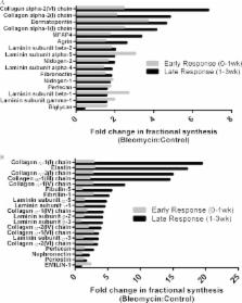
          Fibrotic disease is characterized by the pathological accumulation of extracellular matrix (ECM) proteins. Surprisingly, very little is known about the synthesis and degradation rates of the many proteins and proteoglycans that constitute healthy or pathological extracellular matrix. A comprehensive understanding of altered ECM protein synthesis and degradation during the onset and progression of fibrotic disease would be immensely valuable. We have developed a dynamic proteomics platform that quantifies the fractional synthesis rates of large numbers of proteins via stable isotope labeling and LC/MS-based mass isotopomer analysis. Here, we present the first broad analysis of ECM protein kinetics during the onset of experimental pulmonary fibrosis. Mice were labeled with heavy water for up to 21 days following the induction of lung fibrosis with bleomycin. Lung tissue was subjected to sequential protein extraction to fractionate cellular, guanidine-soluble ECM proteins and residual insoluble ECM proteins. Fractional synthesis rates were calculated for 34 ECM proteins or protein subunits, including collagens, proteoglycans, and microfibrillar proteins. Overall, fractional synthesis rates of guanidine-soluble ECM proteins were faster than those of insoluble ECM proteins, suggesting that the insoluble fraction reflected older, more mature matrix components. This was confirmed through the quantitation of pyridinoline cross-links in each protein fraction. In fibrotic lung tissue, there was a significant increase in the fractional synthesis of unique sets of matrix proteins during early (pre-1 week) and late (post-1 week) fibrotic response. Furthermore, we isolated fast turnover subpopulations of several ECM proteins ( e.g. type I collagen) based on guanidine solubility, allowing for accelerated detection of increased synthesis of typically slow-turnover protein populations. This establishes the presence of multiple kinetic pools of pulmonary collagen in vivo with altered turnover rates during evolving fibrosis. These data demonstrate the utility of dynamic proteomics in analyzing changes in ECM protein turnover associated with the onset and progression of fibrotic disease.

          Related collections

          Most cited references29

          • Record: found
          • Abstract: found
          • Article: not found

          An overview of tissue and whole organ decellularization processes.

          Biologic scaffold materials composed of extracellular matrix (ECM) are typically derived by processes that involve decellularization of tissues or organs. Preservation of the complex composition and three-dimensional ultrastructure of the ECM is highly desirable but it is recognized that all methods of decellularization result in disruption of the architecture and potential loss of surface structure and composition. Physical methods and chemical and biologic agents are used in combination to lyse cells, followed by rinsing to remove cell remnants. Effective decellularization methodology is dictated by factors such as tissue density and organization, geometric and biologic properties desired for the end product, and the targeted clinical application. Tissue decellularization with preservation of ECM integrity and bioactivity can be optimized by making educated decisions regarding the agents and techniques utilized during processing. An overview of decellularization methods, their effect upon resulting ECM structure and composition, and recently described perfusion techniques for whole organ decellularization techniques are presented herein. Copyright © 2011 Elsevier Ltd. All rights reserved.
            Bookmark
            • Record: found
            • Abstract: found
            • Article: not found

            Acellular normal and fibrotic human lung matrices as a culture system for in vitro investigation.

            Extracellular matrix (ECM) is a dynamic tissue that contributes to organ integrity and function, and its regulation of cell phenotype is a major aspect of cell biology. However, standard in vitro culture approaches are of unclear physiologic relevance because they do not mimic the compositional, architectural, or distensible nature of a living organ. In the lung, fibroblasts exist in ECM-rich interstitial spaces and are key effectors of lung fibrogenesis. To better address how ECM influences fibroblast phenotype in a disease-specific manner, we developed a culture system using acellular human normal and fibrotic lungs. Decellularization was achieved using treatment with detergents, salts, and DNase. The resultant matrices can be sectioned as uniform slices within which cells were cultured. We report that the decellularization process effectively removes cellular and nuclear material while retaining native dimensionality and stiffness of lung tissue. We demonstrate that lung fibroblasts reseeded into acellular lung matrices can be subsequently assayed using conventional protocols; in this manner we show that fibrotic matrices clearly promote transforming growth factor-β-independent myofibroblast differentiation compared with normal matrices. Furthermore, comprehensive analysis of acellular matrix ECM details significant compositional differences between normal and fibrotic lungs, paving the way for further study of novel hypotheses. This methodology is expected to allow investigation of important ECM-based hypotheses in human tissues and permits future scientific exploration in an organ- and disease-specific manner.
              Bookmark
              • Record: found
              • Abstract: found
              • Article: not found

              Pulmonary fibrosis: patterns and perpetrators.

              Pulmonary fibrosis occurs in a variety of clinical settings, constitutes a major cause of morbidity and mortality, and represents an enormous unmet medical need. However, the disease is heterogeneous, and the failure to accurately discern between forms of fibrosing lung diseases leads to inaccurate treatments. Pulmonary fibrosis occurring in the context of connective tissue diseases is often characterized by a distinct pattern of tissue pathology and may be amenable to immunosuppressive therapies. In contrast, idiopathic pulmonary fibrosis (IPF) is a progressive and lethal form of fibrosing lung disease that is recalcitrant to therapies that target the immune system. Although animal models of fibrosis imperfectly recapitulate IPF, they have yielded numerous targets for therapeutic intervention. Understanding the heterogeneity of these diseases and elucidating the final common pathways of fibrogenesis are critical for the development of efficacious therapies for severe fibrosing lung diseases.
                Bookmark

                Author and article information

                Journal
                Mol Cell Proteomics
                Mol. Cell Proteomics
                mcprot
                mcprot
                MCP
                Molecular & Cellular Proteomics : MCP
                The American Society for Biochemistry and Molecular Biology
                1535-9476
                1535-9484
                July 2014
                16 April 2014
                : 13
                : 7
                : 1741-1752
                Affiliations
                [1]From *KineMed Inc., 5980 Horton St., Suite 470, Emeryville California 94608;
                [2]§Department of Nutritional Science and Toxicology, University of California, Berkeley, Berkeley, California 94720
                Author notes
                ‡ To whom correspondence should be addressed: Martin L. Decaris, KineMed Inc., 5980 Horton St., Suite 470, Emeryville, CA 94608, Tel.: 510-655-6525, Fax: 510-655-6506, E-mail: mdecaris@ 123456kinemed.com .
                Article
                M113.037267
                10.1074/mcp.M113.037267
                4083112
                24741116
                df93b1d5-d14d-47cb-8df3-5e89fb5fbcb6
                © 2014 by The American Society for Biochemistry and Molecular Biology, Inc.

                Author's Choice—Final version full access.

                History
                : 17 December 2013
                : 9 April 2014
                Categories
                Research

                Molecular biology
                Molecular biology

                Comments

                Comment on this article