71
views
0
recommends
+1 Recommend
0 collections
    0
    shares
      • Record: found
      • Abstract: found
      • Article: found
      Is Open Access

      A method for electrophysiological characterization of hamster retinal ganglion cells using a high-density CMOS microelectrode array

      research-article

      Read this article at

      Bookmark
          There is no author summary for this article yet. Authors can add summaries to their articles on ScienceOpen to make them more accessible to a non-specialist audience.

          Abstract

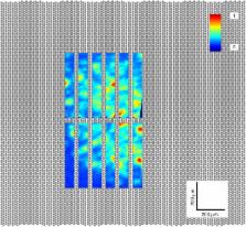
          Knowledge of neuronal cell types in the mammalian retina is important for the understanding of human retinal disease and the advancement of sight-restoring technology, such as retinal prosthetic devices. A somewhat less utilized animal model for retinal research is the hamster, which has a visual system that is characterized by an area centralis and a wide visual field with a broad binocular component. The hamster retina is optimally suited for recording on the microelectrode array (MEA), because it intrinsically lies flat on the MEA surface and yields robust, large-amplitude signals. However, information in the literature about hamster retinal ganglion cell functional types is scarce. The goal of our work is to develop a method featuring a high-density (HD) complementary metal-oxide-semiconductor (CMOS) MEA technology along with a sequence of standardized visual stimuli in order to categorize ganglion cells in isolated Syrian Hamster ( Mesocricetus auratus) retina. Since the HD-MEA is capable of recording at a higher spatial resolution than most MEA systems (17.5 μm electrode pitch), we were able to record from a large proportion of RGCs within a selected region. Secondly, we chose our stimuli so that they could be run during the experiment without intervention or computation steps. The visual stimulus set was designed to activate the receptive fields of most ganglion cells in parallel and to incorporate various visual features to which different cell types respond uniquely. Based on the ganglion cell responses, basic cell properties were determined: direction selectivity, speed tuning, width tuning, transience, and latency. These properties were clustered to identify ganglion cell types in the hamster retina. Ultimately, we recorded up to a cell density of 2780 cells/mm 2 at 2 mm (42°) from the optic nerve head. Using five parameters extracted from the responses to visual stimuli, we obtained seven ganglion cell types.

          Related collections

          Most cited references60

          • Record: found
          • Abstract: not found
          • Article: not found

          Silhouettes: A graphical aid to the interpretation and validation of cluster analysis

            Bookmark
            • Record: found
            • Abstract: found
            • Article: not found

            The types of retinal ganglion cells: current status and implications for neuronal classification.

            In the retina, photoreceptors pass visual information to interneurons, which process it and pass it to retinal ganglion cells (RGCs). Axons of RGCs then travel through the optic nerve, telling the rest of the brain all it will ever know about the visual world. Research over the past several decades has made clear that most RGCs are not merely light detectors, but rather feature detectors, which send a diverse set of parallel, highly processed images of the world on to higher centers. Here, we review progress in classification of RGCs by physiological, morphological, and molecular criteria, making a particular effort to distinguish those cell types that are definitive from those for which information is partial. We focus on the mouse, in which molecular and genetic methods are most advanced. We argue that there are around 30 RGC types and that we can now account for well over half of all RGCs. We also use RGCs to examine the general problem of neuronal classification, arguing that insights and methods from the retina can guide the classification enterprise in other brain regions.
              Bookmark
              • Record: found
              • Abstract: found
              • Article: not found

              A review of methods for spike sorting: the detection and classification of neural action potentials.

              The detection of neural spike activity is a technical challenge that is a prerequisite for studying many types of brain function. Measuring the activity of individual neurons accurately can be difficult due to large amounts of background noise and the difficulty in distinguishing the action potentials of one neuron from those of others in the local area. This article reviews algorithms and methods for detecting and classifying action potentials, a problem commonly referred to as spike sorting. The article first discusses the challenges of measuring neural activity and the basic issues of signal detection and classification. It reviews and illustrates algorithms and techniques that have been applied to many of the problems in spike sorting and discusses the advantages and limitations of each and the applicability of these methods for different types of experimental demands. The article is written both for the physiologist wanting to use simple methods that will improve experimental yield and minimize the selection biases of traditional techniques and for those who want to apply or extend more sophisticated algorithms to meet new experimental challenges.
                Bookmark

                Author and article information

                Contributors
                Journal
                Front Neurosci
                Front Neurosci
                Front. Neurosci.
                Frontiers in Neuroscience
                Frontiers Media S.A.
                1662-4548
                1662-453X
                13 October 2015
                2015
                : 9
                : 360
                Affiliations
                [1] 1Bio Engineering Laboratory, Department of Biosystems Science and Engineering, ETH Zurich Basel, Switzerland
                [2] 2Visual Circuits Laboratory, Neuroelectronics Research Flanders Leuven, Belgium
                [3] 3NERF, Imec Leuven, Belgium
                [4] 4Department of Biology, KU Leuven Leuven, Belgium
                Author notes

                Edited by: Michele Giugliano, University of Antwerp, Belgium

                Reviewed by: Axel Blau, Fondazione Istituto Italiano di Tecnologia, Italy; Emiliano Brunamonti, University of Rome Sapienza, Italy

                *Correspondence: Ian L. Jones, Bio Engineering Laboratory, Department Biosystems Science and Engineering, ETH Zurich, WRO 1058, Mattenstrasse 26, 4058 Basel, Switzerland ian.jones@ 123456bsse.ethz.ch

                This article was submitted to Neural Technology, a section of the journal Frontiers in Neuroscience

                Article
                10.3389/fnins.2015.00360
                4602149
                26528115
                e00febd6-6f1a-47d1-a011-fa4076f48650
                Copyright © 2015 Jones, Russell, Farrow, Fiscella, Franke, Müller, Jäckel and Hierlemann.

                This is an open-access article distributed under the terms of the Creative Commons Attribution License (CC BY). The use, distribution or reproduction in other forums is permitted, provided the original author(s) or licensor are credited and that the original publication in this journal is cited, in accordance with accepted academic practice. No use, distribution or reproduction is permitted which does not comply with these terms.

                History
                : 13 July 2015
                : 18 September 2015
                Page count
                Figures: 10, Tables: 0, Equations: 5, References: 72, Pages: 16, Words: 10786
                Funding
                Funded by: European Research Council 10.13039/501100000781
                Award ID: AdG 267351
                Funded by: Schweizerischer Nationalfonds zur Förderung der Wissenschaftlichen Forschung 10.13039/501100001711
                Award ID: CRSII3_141801
                Categories
                Neuroscience
                Methods

                Neurosciences
                retinal ganglion cells,rgc,mea,cmos,visual stimulation,cell classification,retina,sensory encoding

                Comments

                Comment on this article