19
views
0
recommends
+1 Recommend
0 collections
    0
    shares
      • Record: found
      • Abstract: found
      • Article: found
      Is Open Access

      A five-year prospective study of spinal radiographic progression and its predictors in men and women with ankylosing spondylitis

      research-article

      Read this article at

      Bookmark
          There is no author summary for this article yet. Authors can add summaries to their articles on ScienceOpen to make them more accessible to a non-specialist audience.

          Abstract

          Background

          Knowledge about predictors of new spinal bone formation in patients with ankylosing spondylitis (AS) is limited. AS-related spinal alterations are more common in men; however, knowledge of whether predictors differ between sexes is lacking. Our objectives were to study spinal radiographic progression in patients with AS and investigate predictors of progression overall and by sex.

          Methods

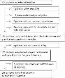
          Swedish patients with AS, age (mean ± SD) 50 ± 13 years, were included in a longitudinal study. At baseline and at 5-year follow up, spinal radiographs were graded according to the modified Stoke Ankylosing Spondylitis Spine Score (mSASSS). Predictors were assessed by questionnaires, spinal mobility tests and blood samples.

          Results

          Of 204 patients included, 166 (81%) were re-examined and 54% were men. Men had significantly higher mean mSASSS at baseline and higher mean increase in mSASSS than women (1.9 ± 2.8 vs. 1.2 ± 3.3; p = 0.005) More men than women developed new syndesmophytes (30% vs. 12%; p = 0.007). Multivariate logistic regression analyses with progression ≥ 2 mSASSS units over 5 years or development of new syndesmophytes as the dependent variable showed that presence of baseline AS-related spinal radiographic alterations and obesity (OR 3.78, 95% CI 1.3 to 11.2) were independent predictors of spinal radiographic progression in both sexes. High C-reactive protein (CRP) was a significant predictor in men, with only a trend seen in women. Smoking predicted progression in men whereas high Bath Ankylosing Spondylitis Metrology Index (BASMI) and exposure to bisphosphonates during follow up (OR 4.78, 95% CI 1.1 to 20.1) predicted progression in women.

          Conclusion

          This first report on sex-specific predictors of spinal radiographic progression shows that predictors may partly differ between the sexes. New predictors identified were obesity in both sexes and exposure to bisphosphonates in women. Among previously known predictors, baseline AS-related spinal radiographic alterations predicted radiographic progression in both sexes, high CRP was a predictor in men (with a trend in women) and smoking was a predictor only in men.

          Trial registration

          ClinicalTrials.gov, NCT00858819. Registered on 9 March 2009. Last updated 28 May 2015.

          Related collections

          Most cited references27

          • Record: found
          • Abstract: found
          • Article: not found

          Assessment of outcome in ankylosing spondylitis: an extended radiographic scoring system.

          To develop and validate an extensive radiographic scoring system for ankylosing spondylitis (AS). The Stoke Ankylosing Spondylitis Spinal Score (SASSS) was modified by adding a score for the cervical spine and defining squaring. This modified SASSS (mSASSS) is the sum of the lumbar and cervical spine score (range 0-72). 370 lateral views of the lumbar and cervical spine were used for development of the mSASSS, standardisation of observers, and for studying reliability. In a 48 week NSAID study of 57 patients, change over time and construct validity were studied. Interobserver correlations of the lumbar and cervical spine scores were good (r>0.95). The interobserver duplicate error was 0.55 in a range from 0 to 36. The mean change in the cervical and lumbar spine scores between weeks 0 and 48 of all patients was 1.45 (range 0-6.0) and 1.06 (0-5.0), respectively (paired t testing, p<0.001). Change in radiological score was seen in 36/57 (63%) patients (lumbar and cervical spine 11, cervical spine 12, lumbar spine 13 patients). The mSASSS is useful for assessing extensive radiographic damage in AS. It is reliable, detects changes over 48 weeks, and shows a satisfactory face and construct validity.
            Bookmark
            • Record: found
            • Abstract: found
            • Article: found
            Is Open Access

            TNF blockers inhibit spinal radiographic progression in ankylosing spondylitis by reducing disease activity: results from the Swiss Clinical Quality Management cohort

            Objectives To analyse the impact of tumour necrosis factor inhibitors (TNFis) on spinal radiographic progression in ankylosing spondylitis (AS). Methods Patients with AS in the Swiss Clinical Quality Management cohort with up to 10 years of follow-up and radiographic assessments every 2 years were included. Radiographs were scored by two readers according to the modified Stoke Ankylosing Spondylitis Spine Score (mSASSS) with known chronology. The relationship between TNFi use before a 2-year radiographic interval and progression within the interval was investigated using binomial generalised estimating equation models with adjustment for potential confounding and multiple imputation of missing values. Ankylosing Spondylitis Disease Activity Score (ASDAS) was regarded as mediating the effect of TNFi on progression and added to the model in a sensitivity analysis. Results A total of 432 patients with AS contributed to data for 616 radiographic intervals. Radiographic progression was defined as an increase in ≥2 mSASSS units in 2 years. Mean (SD) mSASSS increase was 0.9 (2.6) units in 2 years. Prior use of TNFi reduced the odds of progression by 50% (OR 0.50, 95% CI 0.28 to 0.88) in the multivariable analysis. While no direct effect of TNFi on progression was present in an analysis including time-varying ASDAS (OR 0.61, 95% CI 0.34 to 1.08), the indirect effect, via a reduction in ASDAS, was statistically significant (OR 0.75, 95% CI 0.59 to 0.97). Conclusion TNFis are associated with a reduction of spinal radiographic progression in patients with AS. This effect seems mediated through the inhibiting effect of TNFi on disease activity.
              Bookmark
              • Record: found
              • Abstract: found
              • Article: not found

              Bone metabolism in obesity and weight loss.

              Excess body weight due to obesity has traditionally been considered to have a positive effect on bone; however, more recent findings suggest that bone quality is compromised. Both obesity and caloric restriction increase fracture risk and are regulated by endocrine factors and cytokines that have direct and indirect effects on bone and calcium absorption. Weight reduction will decrease bone mass and mineral density, but this varies by the individual's age, gender, and adiposity. Dietary modifications, exercise, and medications have been shown to attenuate the bone loss associated with weight reduction. Future obesity and weight loss trials would benefit from assessment of key hormones, adipokine and gut peptides that regulate calcium absorption, and bone mineral density and quality by using sensitive techniques in high-risk populations.
                Bookmark

                Author and article information

                Contributors
                +46(0)31-3428954 , anna.deminger@vgregion.se
                eva.klingberg@vgregion.se
                mats.geijer@med.lu.se
                jan.gothlin@gmail.com
                martin.hedberg@vgregion.se
                eva.rehnberg@vgregion.se
                hans.carlsten@rheuma.gu.se
                lennart.jacobsson@gu.se
                helena.forsblad@rheuma.gu.se
                Journal
                Arthritis Res Ther
                Arthritis Res. Ther
                Arthritis Research & Therapy
                BioMed Central (London )
                1478-6354
                1478-6362
                3 August 2018
                3 August 2018
                2018
                : 20
                : 162
                Affiliations
                [1 ]ISNI 0000 0000 9919 9582, GRID grid.8761.8, Department of Rheumatology and Inflammation Research, , Sahlgrenska Academy at University of Gothenburg, ; Box 480, 405 30 Gothenburg, Sweden
                [2 ]GRID grid.411843.b, Department of Radiology, , Skåne University Hospital, ; 221 85 Lund, Sweden
                [3 ]ISNI 0000 0001 0930 2361, GRID grid.4514.4, Faculty of Medicine, , Lund University, ; Box 117, 221 00 Lund, Sweden
                [4 ]ISNI 000000009445082X, GRID grid.1649.a, Department of Radiology, , Sahlgrenska University Hospital, ; Mölndal, 431 80 Mölndal, Sweden
                [5 ]ISNI 0000 0004 0624 0304, GRID grid.468026.e, Section of Rheumatology, , Södra Älvsborg Hospital, ; 501 82 Borås, Sweden
                [6 ]Section of Rheumatology, Alingsås Hospital, 441 33 Alingsås, Sweden
                [7 ]ISNI 0000 0001 1034 3451, GRID grid.12650.30, Department of Public Health and Clinical Medicine, Rheumatology, , 901 87 Umeå University, ; Umeå, Sweden
                Author information
                http://orcid.org/0000-0001-6423-1623
                Article
                1665
                10.1186/s13075-018-1665-1
                6091099
                30075808
                e13637ef-3166-4100-bd23-2f63509bdac0
                © The Author(s). 2018

                Open AccessThis article is distributed under the terms of the Creative Commons Attribution 4.0 International License ( http://creativecommons.org/licenses/by/4.0/), which permits unrestricted use, distribution, and reproduction in any medium, provided you give appropriate credit to the original author(s) and the source, provide a link to the Creative Commons license, and indicate if changes were made. The Creative Commons Public Domain Dedication waiver ( http://creativecommons.org/publicdomain/zero/1.0/) applies to the data made available in this article, unless otherwise stated.

                History
                : 23 February 2018
                : 10 July 2018
                Funding
                Funded by: The Health and Medical Care Executive Board of the Västra Götaland
                Funded by: FundRef http://dx.doi.org/10.13039/100008448, Rune och Ulla Amlövs Stiftelse för Neurologisk och Reumatologisk Forskning;
                Funded by: The Swedish Rheumatism Association
                Funded by: FundRef http://dx.doi.org/10.13039/501100008106, Reumatikerdistriktet i Göteborg;
                Funded by: FundRef http://dx.doi.org/10.13039/501100005689, Göteborgs Läkaresällskap;
                Funded by: FundRef http://dx.doi.org/10.13039/501100007687, Svenska Läkaresällskapet;
                Funded by: Västra Götalandsregionen (SE) (agreement concerning research and education of doctors)
                Funded by: Controlling Chronic Inflammatory Diseases with Combined Efforts (COMBINE)
                Funded by: FundRef http://dx.doi.org/10.13039/501100008090, Stiftelsen Reuma Forskningsfond Margareta;
                Categories
                Research Article
                Custom metadata
                © The Author(s) 2018

                Orthopedics
                ankylosing spondylitis,outcomes research,treatment,inflammation,longitudinal study,radiography

                Comments

                Comment on this article