23
views
0
recommends
+1 Recommend
0 collections
    0
    shares
      • Record: found
      • Abstract: found
      • Article: found
      Is Open Access

      Neutrophil elastase inhibition effectively rescued angiopoietin-1 decrease and inhibits glial scar after spinal cord injury

      research-article

      Read this article at

      Bookmark
          There is no author summary for this article yet. Authors can add summaries to their articles on ScienceOpen to make them more accessible to a non-specialist audience.

          Abstract

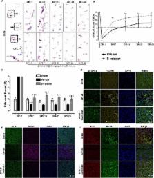
          After spinal cord injury (SCI), neutrophil elastase (NE) released at injury site disrupts vascular endothelium integrity and stabilization. Angiopoietins (ANGPTs) are vascular growth factors that play an important role in vascular stabilization. We hypothesized that neutrophil elastase is one of the key determinants of vascular endothelium disruption/destabilization and affects angiopoietins expression after spinal cord injury. To test this, tubule formation and angiopoietins expression were assessed in endothelial cells exposed to different concentrations of recombinant neutropil elastase. Then, the expression of angiopoietin-1, angiopoietin-2, and neutrophil elastase was determined at 3 h and at 1, 3, 5, 7, 14, 21, and 28 days in a clinically relevant model of moderate compression (35 g for 5 min at T10) spinal cord injury. A dichotomy between the levels of angiopoietin-1 and angiopoietin-2 was observed; thus, we utilized a specific neutrophil elastase inhibitor (sivelestat sodium; 30 mg/kg, i.p., b.i.d.) after spinal cord injury. The expression levels of neutropil elastase and angiopoietin-2 increased, and that of angiopoietin-1 decreased after spinal cord injury in rats. The sivelestat regimen, optimized via a pharmacokinetics study, had potent effects on vascular stabilization by upregulating angiopoietin-1 via the AKT pathway and preventing tight junction protein degradation. Moreover, sivelestat attenuated the levels of inflammatory cytokines and chemokines after spinal cord injury and hence subsequently alleviated secondary damage observed as a reduction in glial scar formation and the promotion of blood vessel formation and stabilization. As a result, hindlimb locomotor function significantly recovered in the sivelestat-treated animals as determined by the Basso, Beattie, and Bresnahan scale and footprint analyses. Furthermore, sivelestat treatment attenuated neuropathic pain as assessed by responses to von Frey filaments after spinal cord injury. Thus, our result suggests that inhibiting neutropil elastase by administration of sivelestat is a promising therapeutic strategy to inhibit glial scar and promote functional recovery by upregulating angiopoietin-1 after spinal cord injury.

          Electronic supplementary material

          The online version of this article (10.1186/s40478-018-0576-3) contains supplementary material, which is available to authorized users.

          Related collections

          Most cited references76

          • Record: found
          • Abstract: found
          • Article: not found

          Endothelial cell-to-cell junctions: molecular organization and role in vascular homeostasis.

          Intercellular junctions mediate adhesion and communication between adjoining endothelial and epithelial cells. In the endothelium, junctional complexes comprise tight junctions, adherens junctions, and gap junctions. The expression and organization of these complexes depend on the type of vessels and the permeability requirements of perfused organs. Gap junctions are communication structures, which allow the passage of small molecular weight solutes between neighboring cells. Tight junctions serve the major functional purpose of providing a "barrier" and a "fence" within the membrane, by regulating paracellular permeability and maintaining cell polarity. Adherens junctions play an important role in contact inhibition of endothelial cell growth, paracellular permeability to circulating leukocytes and solutes. In addition, they are required for a correct organization of new vessels in angiogenesis. Extensive research in the past decade has identified several molecular components of the tight and adherens junctions, including integral membrane and intracellular proteins. These proteins interact both among themselves and with other molecules. Here, we review the individual molecules of junctions and their complex network of interactions. We also emphasize how the molecular architectures and interactions may represent a mechanistic basis for the function and regulation of junctions, focusing on junction assembly and permeability regulation. Finally, we analyze in vivo studies and highlight information that specifically relates to the role of junctions in vascular endothelial cells.
            Bookmark
            • Record: found
            • Abstract: found
            • Article: not found

            Angiopoietin-2 sensitizes endothelial cells to TNF-alpha and has a crucial role in the induction of inflammation.

            The angiopoietins Ang-1 and Ang-2 have been identified as ligands of the receptor tyrosine kinase Tie-2 (refs. 1,2). Paracrine Ang-1-mediated activation of Tie-2 acts as a regulator of vessel maturation and vascular quiescence. In turn, the antagonistic ligand Ang-2 acts by an autocrine mechanism and is stored in endothelial Weibel-Palade bodies from where it can be rapidly released upon stimulation. The rapid release of Ang-2 implies functions of the angiopoietin-Tie system beyond its established role during vascular morphogenesis as a regulator of rapid vascular responses. Here we show that mice deficient in Ang-2 (encoded by the gene Angpt2) cannot elicit an inflammatory response in thioglycollate-induced or Staphylococcus aureus-induced peritonitis, or in the dorsal skinfold chamber model. Recombinant Ang-2 restores the inflammation defect in Angpt2(-/-) mice. Intravital microscopy showed normal TNF-alpha-induced leukocyte rolling in the vasculature of Angpt2(-/-)mice, but rolling cells did not firmly adhere to activated endothelium. Cellular experiments showed that Ang-2 promotes adhesion by sensitizing endothelial cells toward TNF-alpha and modulating TNF-alpha-induced expression of endothelial cell adhesion molecules. Together, these findings identify Ang-2 as an autocrine regulator of endothelial cell inflammatory responses. Ang-2 thereby acts as a switch of vascular responsiveness exerting a permissive role for the activities of proinflammatory cytokines.
              Bookmark
              • Record: found
              • Abstract: found
              • Article: not found

              Management of Sepsis

              New England Journal of Medicine, 355(16), 1699-1713
                Bookmark

                Author and article information

                Contributors
                +86-20-8733-0098-803 , zengx33@mail.sysu.edu.cn
                +82-10-7124-2571 , hanib@cha.ac.kr
                Journal
                Acta Neuropathol Commun
                Acta Neuropathol Commun
                Acta Neuropathologica Communications
                BioMed Central (London )
                2051-5960
                7 August 2018
                7 August 2018
                2018
                : 6
                : 73
                Affiliations
                [1 ]ISNI 0000 0004 0570 1076, GRID grid.452398.1, Department of Neurosurgery, , CHA University School of Medicine, CHA Bundang Medical Center, ; Seongnam-si, Gyeonggi-do 13496 Republic of Korea
                [2 ]ISNI 0000 0004 0647 3511, GRID grid.410886.3, Department of Biomedical Science, , CHA University, ; Seongnam-si, Gyeonggi-do Republic of Korea
                [3 ]ISNI 0000000417581884, GRID grid.18887.3e, Molecular Neurobiology Laboratory, Division of Neuroscience, , San Raffaele Scientific Institute, ; Milan, Italy
                [4 ]ISNI 0000 0001 2171 7818, GRID grid.289247.2, Department of Neurosurgery, , Kyung Hee University, ; Dongdaemun-gu, Seoul 02447 Republic of Korea
                [5 ]ISNI 0000 0004 0647 3511, GRID grid.410886.3, College of Pharmacy, , CHA University, ; Seongnam-si, Gyeonggi-do Republic of Korea
                [6 ]Department of Neurosurgery, Kyungpook National University Hospital, Kyungpook National University, 130, Dongdeok-ro, Jung-gu, Daegu, 41944 Republic of Korea
                [7 ]ISNI 0000 0001 0661 1556, GRID grid.258803.4, Department of Neurosurgery, , School of Medicine,Kyungpook National University, ; 130, Dongdeok-ro, Jung-gu, Daegu, 41944 Republic of Korea
                [8 ]ISNI 0000 0001 2360 039X, GRID grid.12981.33, Department of Histology and Embryology, , Zhongshan School of Medicine, Sun Yat-sen University, ; Guangzhou, 510080 Guangdong Province China
                Article
                576
                10.1186/s40478-018-0576-3
                6080383
                30086801
                e14cf76a-4b73-4204-acdd-3c4651f1f31c
                © The Author(s). 2018

                Open AccessThis article is distributed under the terms of the Creative Commons Attribution 4.0 International License ( http://creativecommons.org/licenses/by/4.0/), which permits unrestricted use, distribution, and reproduction in any medium, provided you give appropriate credit to the original author(s) and the source, provide a link to the Creative Commons license, and indicate if changes were made. The Creative Commons Public Domain Dedication waiver ( http://creativecommons.org/publicdomain/zero/1.0/) applies to the data made available in this article, unless otherwise stated.

                History
                : 25 June 2018
                : 23 July 2018
                Funding
                Funded by: FundRef http://dx.doi.org/10.13039/501100003725, National Research Foundation of Korea;
                Award ID: NRF-2015H1D3A1066543
                Award ID: NRF-2017R1C1B2011772
                Award Recipient :
                Funded by: Korea Healthcare Technology Research & Development Project
                Award ID: HI16C1559
                Funded by: FundRef http://dx.doi.org/10.13039/501100001809, National Natural Science Foundation of China;
                Award ID: 31600780
                Award Recipient :
                Categories
                Research
                Custom metadata
                © The Author(s) 2018

                neutrophil elastase,spinal cord injury,glial scar,angiopoietins,functional recovery,neuropathic pain

                Comments

                Comment on this article