Inviting an author to review:
Find an author and click ‘Invite to review selected article’ near their name.
Search for authorsSearch for similar articles
26
views
0
recommends
+1 Recommend
0 collections
    0
    shares
      • Record: found
      • Abstract: found
      • Article: found
      Is Open Access

      Comparison of cadaveric and isomorphic virtual haptic simulation in temporal bone training

      research-article

      Read this article at

      Bookmark
          There is no author summary for this article yet. Authors can add summaries to their articles on ScienceOpen to make them more accessible to a non-specialist audience.

          Abstract

          Background

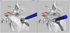
          Virtual surgery may improve learning and provides an opportunity for pre-operative surgical rehearsal. We describe a novel haptic temporal bone simulator specifically developed for multicore processing and improved visual realism. A position locking algorithm for enhanced drill-bone interaction and haptic fidelity is further employed. The simulation construct is evaluated against cadaveric education.

          Methods

          A voxel-based simulator was designed for multicore architecture employing Marching Cubes and Laplacian smoothing to perform real-time haptic and graphic rendering of virtual bone.

          Ten Otolaryngology trainees dissected a cadaveric temporal bone (CTB) followed by a virtual isomorphic haptic model (VM) based on derivative microCT data. Participants rated 1) physical characteristics, 2) specific anatomic constructs, 3) usefulness in skill development and 4) perceived educational value. The survey instrument employed a Likert scale (1-7).

          Results

          Residents were equivocal about the physical properties of the VM, as cortical (3.2 ± 2.0) and trabecular (2.8 ± 1.6) bone drilling character was appraised as dissimilar to CTB. Overall similarity to cadaveric training was moderate (3.5 ± 1.8). Residents generally felt the VM was beneficial in skill development, rating it highest for translabyrinthine skull-base approaches (5.2 ± 1.3). The VM was considered an effective (5.4 ± 1.5) and accurate (5.7 ± 1.4) training tool which should be integrated into resident education (5.5 ± 1.4). The VM was thought to improve performance (5.3 ± 1.8) and confidence (5.3 ± 1.9) and was highly rated for anatomic learning (6.1 ± 1.9).

          Conclusion

          Study participants found the VM to be a beneficial and effective platform for learning temporal bone anatomy and surgical techniques. They identify some concern with limited physical realism likely owing to the haptic device interface. This study is the first to compare isomorphic simulation in education. This significantly removes possible confounding features as the haptic simulation was based on derivative imaging.

          Related collections

          Most cited references23

          • Record: found
          • Abstract: not found
          • Article: not found

          Improved Laplacian Smoothing of Noisy Surface Meshes

            Bookmark
            • Record: found
            • Abstract: found
            • Article: not found

            Generation of a 3D printed temporal bone model with internal fidelity and validation of the mechanical construct.

            To generate a rapid-prototyped temporal bone model from computed tomography (CT) data with a specific focus on internal anatomic fidelity. Research ethics board-approved prospective cohort study. Current iterations of a rapid-prototyped temporal bone model are complicated by absent void spaces and inconsistent bone density due to limited infiltrant exposure. The creation of a high-fidelity model allows surgical trainees to practice in a standardized and reproducible training environment. This learning paradigm will significantly augment resident understanding of surgical approaches and techniques to prevent adverse outcomes. We describe a technique for generating internally accurate rapid-prototyped anatomical models with solid and hollow structures, including void spaces. The novel slicing algorithm digitally deconstructs a model into segments and permits removal of extraneous print material and allows infiltrant penetration of the entire bone structure. Precise reassembly is facilitated by digitally generated fiducials. Infiltrant of choice was determined by expert assessment and subjected to objective mechanical property assessment with comparison to cadaveric sheep bone. The printed bone models are highly realistic. Void space representation was excellent with 88% concordance between cadaveric bone and the resultant rapid-prototyped temporal bone model. Ultimately, cyanoacrylate with hydroquinone was determined to be the most appropriate infiltrant for both cortical and trabecular simulation. The mechanical properties of all tested infiltrants were similar to real bone. This model serves as an excellent replica of a human temporal bone for training and preoperative surgical rehearsal and can be dissected in a true-to-life fashion.
              Bookmark
              • Record: found
              • Abstract: found
              • Article: not found

              Review of temporal bone dissection teaching: how it was, is and will be.

              We aimed to review the history of anatomical dissection, and to examine how modern educational techniques will change the way temporal bone dissection is taught to otolaryngology trainees. Review of the literature using Medline, Embase and PubMed database searches. Temporal bone anatomy has traditionally been taught using cadaveric specimens. However, resources such as three-dimensional reconstructed models and 'virtual reality' temporal bone simulators have a place in educating the otolaryngology trainee. We should encourage the use of fresh frozen cadaveric temporal bone specimens for future otologists. Artificial three-dimensional models and virtual reality temporal bone simulators can be used to educate junior trainees, thus conserving the scarce resource of cadaveric bones.
                Bookmark

                Author and article information

                Contributors
                danawong8@gmail.com
                bertram.j.unger@gmail.com
                jaykraut@gmail.com
                jpisa@hsc.mb.ca
                charlotte_abady@hotmail.com
                jordanhochman@hotmail.com
                Journal
                J Otolaryngol Head Neck Surg
                J Otolaryngol Head Neck Surg
                Journal of Otolaryngology - Head & Neck Surgery
                BioMed Central (London )
                1916-0208
                1916-0216
                13 October 2014
                13 October 2014
                2014
                : 43
                : 1
                : 31
                Affiliations
                [ ]Department of Otolaryngology Head and Neck Surgery, University of Manitoba, Winnipeg, Manitoba Canada
                [ ]Clinical Learning and Simulation Facility, Department of Medical Education, Faculty of Medicine, University of Manitoba, Winnipeg, Manitoba Canada
                [ ]Department of Medical Education, Faculty of Medicine, University of Manitoba, Winnipeg, Manitoba Canada
                [ ]Surgical Hearing Implant Program, Department of Otolaryngology - Head and Neck Surgery, University of Manitoba, GB421, 820 Sherbrook Street, Winnipeg, Manitoba R3A 1R9 Canada
                [ ]Neurotologic Surgery, Department of Otolaryngology Head and Neck Surgery, Faculty of Medicine, University of Manitoba, GB421, 820 Sherbrook Street, Winnipeg, Manitoba R3A 1R9 Canada
                Article
                31
                10.1186/s40463-014-0031-9
                4198678
                25312968
                e23c4455-bf44-4611-b9d4-9e3eab3caa3e
                © Wong et al.; licensee BioMed Central Ltd. 2014

                This is an Open Access article distributed under the terms of the Creative Commons Attribution License ( http://creativecommons.org/licenses/by/4.0), which permits unrestricted use, distribution, and reproduction in any medium, provided the original work is properly credited. The Creative Commons Public Domain Dedication waiver ( http://creativecommons.org/publicdomain/zero/1.0/) applies to the data made available in this article, unless otherwise stated.

                History
                : 27 May 2014
                : 18 July 2014
                Categories
                Original Research Article
                Custom metadata
                © The Author(s) 2014

                medical simulation,haptic,real time marching cubes,temporal bone

                Comments

                Comment on this article