10
views
0
recommends
+1 Recommend
1 collections
    0
    shares
      • Record: found
      • Abstract: found
      • Article: found
      Is Open Access

      Comparison of inflammatory markers between brucella and non-brucella epididymo-orchitis

      research-article

      Read this article at

      ScienceOpenPublisherPMC
      Bookmark
          There is no author summary for this article yet. Authors can add summaries to their articles on ScienceOpen to make them more accessible to a non-specialist audience.

          ABSTRACT

          Objectives:

          Brucellosis is a multi-system infectious disease that is associated with inflammation, which causes an increase in acute phase reactants. Hematological inflammatory markers of brucellosis include mean platelet volume (MPV), red cell distribution width (RDW), neutrophil/lymphocyte ratio (NLR), and platelet/lymphocyte ratio (PLR). In this study, we aimed to evaluate the diagnostic value of hematological inflammatory markers in Brucella epididymo-orchitis (BEO), and to investigate the utility of these markers for differential diagnosis from non-Brucella epididymo-orchitis (non-BEO).

          Materials and Methods:

          We retrospectively reviewed the records of 22 BEO and 50 non-BEO patients. Hematological parameters were recorded and compared between the two groups. The main diagnostic criteria for BEO were positive clinical findings (i.e., testicular pain, tenderness and scrotal swelling), a positive Rose Bengal test result, standard tube agglutination (STA) titer ≥ 1/160, and/or a positive blood culture.

          Results:

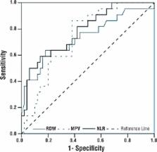
          The most decisive factors in discriminating between BEO and non-BEO were NLR, RDW, and MPV, in decreasing order of their strength. Regardless of other factors, NLR values < 2.3 significantly increased the odds of BEO (OR=8.080, 95% CI: 1.929-33.843, p=0.004). After adjusting for other factors, RDW values >14.45% significantly increased the odds of BEO (OR=7.020, 95% CI: 1.749-28.176, p=0.006). Independent of the other factors, patients with MPV < 7.65 fL had a 6.336 times higher risk for BEO (95% CI: 1.393 - 28.822, p=0.017).

          Conclusion:

          Hematological inflammatory markers such as NLR, RDW, and MPV can aid in the differential diagnosis of BEO and non-BEO.

          Related collections

          Most cited references36

          • Record: found
          • Abstract: found
          • Article: found
          Is Open Access

          Preoperative platelet lymphocyte ratio (PLR) is superior to neutrophil lymphocyte ratio (NLR) as a predictive factor in patients with esophageal squamous cell carcinoma

          Background Recent studies have shown that the presence of systemic inflammation correlates with poor survival in various cancers. The aim of this study was to determinate the prognostic value of the neutrophil lymphocyte ratio (NLR) and the platelet lymphocyte ratio (PLR) in patients with esophageal squamous cell carcinoma (ESCC). Methods Preoperative NLR and PLR were evaluated in 483 patients undergoing esophagectomy for ESCC from January 2005 to December 2008. The prognostic significance of both markers was then determined by both uni- and multivariate analytical methods. Receiver operating characteristic (ROC) curves were also plotted to verify the accuracy of NLR and PLR for survival prediction. Results High preoperative NLR (≥3.5 versus < 3.5, P = 0.039) and PLR (≥150 versus < 150, P < 0.001) were significantly associated with poor overall survival in multivariate analysis. However, our study demonstrated a better discrimination for the PLR in terms of hazard ratio(HR) than the NLR (HR = 1.840 versus HR = 1.339). Patients with NLR ≥3.5 had significantly poorer overall survival compared to NLR <3.5 (35.4% versus 57.7%, P < 0.001). Patients with PLR ≥150 also had significantly poorer overall survival compared to patients with PLR <150 (32.7% versus 63.5%, P < 0.001). The area under the curve (AUC) was 0.658 (95% confidence interval (CI): 0.610 to 0.706, P < 0.001) for NLR and 0.708 (95% CI: 0.662 to 0.754, P < 0.001) for PLR, indicating that PLR was superior to NLR as a predictive factor in ESCC. Conclusions Preoperative NLR and PLR were significant predictors of overall survival in patients with ESCC. However, PLR is superior to NLR as a predictive factor in patients with ESCC.
            Bookmark
            • Record: found
            • Abstract: found
            • Article: not found

            Complications associated with Brucella melitensis infection: a study of 530 cases.

            We carried out a prospective study of 530 patients older than 14 years of age with brucellosis. We describe the incidence and clinical features of the focal forms of the disease, analyzing some of the possible factors associated with their appearance. One hundred sixty-nine patients (31.9%) had a focal form or complication. Osteoarticular complications were the most frequent, totaling 113 cases (66%), followed by genitourinary with 18 cases (5.1% of males), hepatic (2.5%), neurologic (1.7%), and heart (1.5%). Nine patients (1.7%) had more than 1 complication. In a multivariate analysis, diagnostic delay greater than 30 days (OR 2.0), ESR > 40 mm/hr (OR 1.9), and levels of alpha-2 globulin > 7.5 g/L (OR 6.8) were statistically significant independent variables associated with the presence of focal forms. Twenty-five patients with complications (14.8%) required surgical treatment. The relapse rate was 3.6% for those patients without complications and 4.1% for patients with focal forms (p > 0.05). However, when therapeutic failure, relapses, and mortality were considered together, the risk of an unfavorable evolution was significantly greater in patients with focal forms (10.6% versus 3.6% in patients without complications; OR 1.9, 95% CI 1.4-7.1, p < 0.005). Given the worse prognosis, knowledge and early diagnosis of the focal forms of B. melitensis infection is especially important.
              Bookmark
              • Record: found
              • Abstract: found
              • Article: not found

              White blood cell counts, leukocyte ratios, and eosinophils as inflammatory markers in patients with coronary artery disease.

              Inflammation is a key feature of atherosclerosis and its clinical manifestations. The leukocyte count has emerged as a marker of inflammation that is widely available in clinical practice. Since inflammation plays a key role in atherosclerosis and its end results, discovering new biomarkers of inflammation becomes important in order to help diagnostic accuracy and provide prognostic information about coronary cardiac disease. In acute coronary syndromes and percutaneous coronary intervention, elevated levels of almost all subtypes of white blood cell counts, including eosinophils, monocytes, neutrophils, and lymphocytes, and neutrophil-lymphocyte ratio and eosinophil-leukocyte ratio constitute independent predictors of adverse outcomes. Eosinophil count and eosinophil-leukocyte ratio, in particular, emerge as novel biomarkers for risk stratification in patients with coronary artery disease. Since the presence of eosinophils denotes hypersensitivity inflammation and hypersensitivity associated with Kounis syndrome, this reality is essential for elucidating the etiology of inflammation in order to consider predictive and preventive measures and to apply the appropriate therapeutic methods.
                Bookmark

                Author and article information

                Journal
                Int Braz J Urol
                Int Braz J Urol
                ibju
                International Brazilian Journal of Urology : official journal of the Brazilian Society of Urology
                Sociedade Brasileira de Urologia
                1677-5538
                1677-6119
                Jul-Aug 2018
                Jul-Aug 2018
                : 44
                : 4
                : 771-778
                Affiliations
                [1 ]Department of Urology, Faculty of Medicine, Adiyaman University, Adiyaman, Turkey
                Author notes
                Correspondence address: Mehmet Ozgur Yucel, MD Adiyaman Universitesi Adiyaman Tip Fak Adiyaman None 02100, Turkey E-mail: mozyucel@ 123456yahoo.com

                CONFLICT OF INTEREST

                None declared.

                Article
                S1677-5538.IBJU.2018.0004.0
                10.1590/S1677-5538.IBJU.2018.0004.0
                6092658
                29697933
                e3c8a65a-08cc-4917-bb2a-ef93f1f6d95a

                This is an Open Access article distributed under the terms of the Creative Commons Attribution License, which permits unrestricted use, distribution, and reproduction in any medium, provided the original work is properly cited.

                History
                : 05 January 2018
                : 26 March 2018
                Page count
                Figures: 4, Tables: 4, Equations: 0, References: 34, Pages: 8
                Categories
                Original Article

                brucella,infectious disease medicine,hematologic neoplasms

                Comments

                Comment on this article