18
views
0
recommends
+1 Recommend
0 collections
    0
    shares
      • Record: found
      • Abstract: found
      • Article: found
      Is Open Access

      The endogenous soluble VEGF receptor-2 isoform suppresses lymph node metastasis in a mouse immunocompetent mammary cancer model

      research-article

      Read this article at

      Bookmark
          There is no author summary for this article yet. Authors can add summaries to their articles on ScienceOpen to make them more accessible to a non-specialist audience.

          Abstract

          Background

          Cancer metastasis contributes significantly to cancer mortality and is facilitated by lymphangiogenesis and angiogenesis. A new splicing variant, endogenous soluble vascular endothelial growth factor receptor-2 (esVEGFR-2) that we recently identified is an endogenous selective inhibitor of lymphangiogenesis. To evaluate the antimetastatic potential of esVEGFR-2, gene therapy with vector expressing esVEGFR-2 (pesVEGFR-2) or endostatin (pEndo) as a positive control was conducted on murine metastatic mammary cancer.

          Methods

          Syngeneic inoculated metastatic mammary cancers received direct intratumoral injection of pesVEGFR-2, pEndo or pVec as control, once a week for six weeks. In vivo gene electrotransfer was performed on the tumors after each injection.

          Results

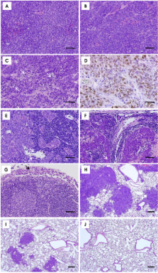
          Deaths from metastasis were much lower in the pesVEGFR-2 and pEndo groups than in those of the pVec. Tumor volume was significantly lower in the pesVEGFR-2 and the pEndo groups throughout the study. Multiplicity of lymph node and lung metastatic nodules was significantly suppressed in the pesVEGFR-2 and pEndo groups. Moreover, the total number of overall metastasis including the other organs was also decreased in these groups. However, pesVEGFR-2 was not able to decrease the number of lungs, ovaries, kidneys and adrenals with metastasis as counted by unilateral or bilateral metastasis. The number of CD34 +/Lyve-1 - blood microvessels was significantly decreased in the pEndo group, while the number of CD34 -/Lyve-1 + lymphatic vessels was significantly decreased in the pesVEGFR-2 and pEndo groups. In addition, a significant reduction in the number of dilated lymphatic vessels containing intraluminal cancer cells was observed in the pesVEGFR-2 and pEndo groups. Levels of apoptosis were significantly increased in the pEndo group, whereas the rates of cell proliferation were significantly decreased in the pesVEGFR-2 and pEndo groups.

          Conclusions

          Our data demonstrate that esVEGFR-2 can inhibit mainly lymph node metastasis. The antimetastatic activity of esVEGFR-2 may be of high clinical significance in the treatment of metastatic breast cancer because lymph node involvement is a most important prognostic factor in cancer patients.

          Related collections

          Most cited references27

          • Record: found
          • Abstract: found
          • Article: not found

          Relation of tumor size, lymph node status, and survival in 24,740 breast cancer cases.

          Two of the most important prognostic indicators for breast cancer are tumor size and extent of axillary lymph node involvement. Data on 24,740 cases recorded in the Surveillance, Epidemiology, and End Results (SEER) Program of the National Cancer Institute were used to evaluate the breast cancer survival experience in a representative sample of women from the United States. Actuarial (life table) methods were used to investigate the 5-year relative survival rates in cases with known operative/pathologic axillary lymph node status and primary tumor diameter. Survival rates varied from 45.5% for tumor diameters equal to or greater than 5 cm with positive axillary nodes to 96.3% for tumors less than 2 cm and with no involved nodes. The relation between tumor size and lymph node status was investigated in detail. Tumor diameter and lymph node status were found to act as independent but additive prognostic indicators. As tumor size increased, survival decreased regardless of lymph node status; and as lymph node involvement increased, survival status also decreased regardless of tumor size. A linear relation was found between tumor diameter and the percent of cases with positive lymph node involvement. The results of our analyses suggest that disease progression to distant sites does not occur exclusively via the axillary lymph nodes, but rather that lymph node status serves as an indicator of the tumor's ability to spread.
            Bookmark
            • Record: found
            • Abstract: found
            • Article: not found

            Vascular endothelial growth factor-C-mediated lymphangiogenesis promotes tumour metastasis.

            Metastasis is a frequent and lethal complication of cancer. Vascular endothelial growth factor-C (VEGF-C) is a recently described lymphangiogenic factor. Increased expression of VEGF-C in primary tumours correlates with dissemination of tumour cells to regional lymph nodes. However, a direct role for VEGF-C in tumour lymphangiogenesis and subsequent metastasis has yet to be demonstrated. Here we report the establishment of transgenic mice in which VEGF-C expression, driven by the rat insulin promoter (Rip), is targeted to beta-cells of the endocrine pancreas. In contrast to wild-type mice, which lack peri-insular lymphatics, RipVEGF-C transgenics develop an extensive network of lymphatics around the islets of Langerhans. These mice were crossed with Rip1Tag2 mice, which develop pancreatic beta-cell tumours that are neither lymphangiogenic nor metastatic. Double-transgenic mice formed tumours surrounded by well developed lymphatics, which frequently contained tumour cell masses of beta-cell origin. These mice frequently developed pancreatic lymph node metastases. Our findings demonstrate that VEGF-C-induced lymphangiogenesis mediates tumour cell dissemination and the formation of lymph node metastases.
              Bookmark
              • Record: found
              • Abstract: found
              • Article: not found

              Biology of vascular endothelial growth factors.

              Angiogenesis is the process by which new blood vessels are formed from existing vessels. The vascular endothelial growth factors (VEGFs) are considered as key molecules in the process of angiogenesis. The VEGF family currently includes VEGF-A, -B, -C, -D, -E, -F and placenta growth factor (PlGF), that bind in a distinct pattern to three structurally related receptor tyrosine kinases, denoted VEGF receptor-1, -2, and -3. VEGF-C and VEGF-D also play a crucial role in the process of lymphangiogenesis. Here, we review the biology of VEGFs and evaluate their role in pathological angiogenesis and lymphangiogenesis.
                Bookmark

                Author and article information

                Journal
                BMC Med
                BMC Medicine
                BioMed Central
                1741-7015
                2010
                3 November 2010
                : 8
                : 69
                Affiliations
                [1 ]Department of Anatomy and Cell Biology, Division of Life Sciences, Osaka Medical College, Osaka, Japan
                [2 ]Department of Ophthalmology and Visual Sciences and Physiology, University of Kentucky, Lexington, KY, USA
                [3 ]Laboratory for Drug Discovery Innovation, Department of Molecular Pharmacology, National Cerebral & Cardiovascular Center Research Institute, Osaka, Japan
                [4 ]Laboratory Animal Center, Osaka Medical College, Osaka, Japan
                Article
                1741-7015-8-69
                10.1186/1741-7015-8-69
                2989928
                21047425
                e54b8393-a6fa-450a-b4ab-1e2f9eee0edf
                Copyright ©2010 Shibata et al; licensee BioMed Central Ltd.

                This is an Open Access article distributed under the terms of the Creative Commons Attribution License ( http://creativecommons.org/licenses/by/2.0), which permits unrestricted use, distribution, and reproduction in any medium, provided the original work is properly cited.

                History
                : 28 July 2010
                : 3 November 2010
                Categories
                Research Article

                Medicine
                Medicine

                Comments

                Comment on this article