48
views
0
recommends
+1 Recommend
1 collections
    0
    shares
      • Record: found
      • Abstract: found
      • Article: found
      Is Open Access

      IMI – Defining and Classifying Myopia: A Proposed Set of Standards for Clinical and Epidemiologic Studies

      research-article

      Read this article at

      ScienceOpenPublisherPMC
      Bookmark
          There is no author summary for this article yet. Authors can add summaries to their articles on ScienceOpen to make them more accessible to a non-specialist audience.

          Abstract

          Purpose

          We provide a standardized set of terminology, definitions, and thresholds of myopia and its main ocular complications.

          Methods

          Critical review of current terminology and choice of myopia thresholds was done to ensure that the proposed standards are appropriate for clinical research purposes, relevant to the underlying biology of myopia, acceptable to researchers in the field, and useful for developing health policy.

          Results

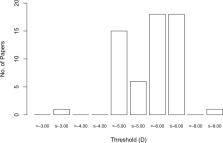
          We recommend that the many descriptive terms of myopia be consolidated into the following descriptive categories: myopia, secondary myopia, axial myopia, and refractive myopia. To provide a framework for research into myopia prevention, the condition of “pre-myopia” is defined. As a quantitative trait, we recommend that myopia be divided into myopia (i.e., all myopia), low myopia, and high myopia. The current consensus threshold value for myopia is a spherical equivalent refractive error ≤ −0.50 diopters (D), but this carries significant risks of classification bias. The current consensus threshold value for high myopia is a spherical equivalent refractive error ≤ −6.00 D. “Pathologic myopia” is proposed as the categorical term for the adverse, structural complications of myopia. A clinical classification is proposed to encompass the scope of such structural complications.

          Conclusions

          Standardized definitions and consistent choice of thresholds are essential elements of evidence-based medicine. It is hoped that these proposals, or derivations from them, will facilitate rigorous, evidence-based approaches to the study and management of myopia.

          Related collections

          Most cited references33

          • Record: found
          • Abstract: found
          • Article: not found

          Effect of Time Spent Outdoors at School on the Development of Myopia Among Children in China: A Randomized Clinical Trial.

          Myopia has reached epidemic levels in parts of East and Southeast Asia. However, there is no effective intervention to prevent the development of myopia.
            Bookmark
            • Record: found
            • Abstract: found
            • Article: found
            Is Open Access

            Global variations and time trends in the prevalence of childhood myopia, a systematic review and quantitative meta-analysis: implications for aetiology and early prevention

            The aim of this review was to quantify the global variation in childhood myopia prevalence over time taking account of demographic and study design factors. A systematic review identified population-based surveys with estimates of childhood myopia prevalence published by February 2015. Multilevel binomial logistic regression of log odds of myopia was used to examine the association with age, gender, urban versus rural setting and survey year, among populations of different ethnic origins, adjusting for study design factors. 143 published articles (42 countries, 374 349 subjects aged 1–18 years, 74 847 myopia cases) were included. Increase in myopia prevalence with age varied by ethnicity. East Asians showed the highest prevalence, reaching 69% (95% credible intervals (CrI) 61% to 77%) at 15 years of age (86% among Singaporean-Chinese). Blacks in Africa had the lowest prevalence; 5.5% at 15 years (95% CrI 3% to 9%). Time trends in myopia prevalence over the last decade were small in whites, increased by 23% in East Asians, with a weaker increase among South Asians. Children from urban environments have 2.6 times the odds of myopia compared with those from rural environments. In whites and East Asians sex differences emerge at about 9 years of age; by late adolescence girls are twice as likely as boys to be myopic. Marked ethnic differences in age-specific prevalence of myopia exist. Rapid increases in myopia prevalence over time, particularly in East Asians, combined with a universally higher risk of myopia in urban settings, suggest that environmental factors play an important role in myopia development, which may offer scope for prevention.
              Bookmark
              • Record: found
              • Abstract: found
              • Article: not found

              Long-term pattern of progression of myopic maculopathy: a natural history study.

              To investigate the long-term progression pattern of myopic maculopathy and to determine the visual prognosis of each progression stage. Retrospective, observational case series. The medical records of 806 eyes of 429 consecutive patients with high myopia (refractive error more than -8.00 diopters [D] or axial length > or =26.5 mm) who were followed for 5-32 years were reviewed. Participants had complete ophthalmological examinations including best-corrected visual acuity, axial length measurements, fluorescein angiography, and color fundus photography, at least once a year. The presence and type of posterior staphyloma was determined by binocular stereoscopic ophthalmoscopy. The types of myopic maculopathy included tessellated fundus, lacquer cracks, diffuse chorioretinal atrophy, patchy chorioretinal atrophy, choroidal neovascularization (CNV), and macular atrophy. None of the patients had received any type of treatment for the maculopathy. The longitudinal long-term progression pattern and the visual prognosis of each type of fundus lesion. During the mean follow-up of 12.7 years, 327 of the 806 highly myopic eyes (40.6%) showed a progression of the myopic maculopathy. The most commonly observed patterns were from tessellated fundus to the development of diffuse atrophy and lacquer cracks, an increase in the width and progression to patchy atrophy in eyes with lacquer cracks, an enlargement of the diffuse atrophy, and the development of patchy atrophy in eyes with diffuse atrophy, and an enlargement and fusion of patches of atrophic areas in eyes with patchy atrophy. Eyes with tessellated fundus, lacquer cracks, diffuse atrophy and patchy atrophy at the initial examination progressed to the development of CNV. Eyes with CNV developed macular atrophy. The fusion of patchy atrophy, the development of CNV, and macular atrophy all led to significant visual decreases. A posterior staphyloma was observed more frequently in eyes that showed progression from tessellated fundus, diffuse atrophy, and patchy atrophy than those without a progression. These findings indicate that myopic maculopathy tends to progress in approximately 40% of highly myopic eyes, and the pattern of progression affects the visual prognosis. Preventive therapy targeting posterior staphyloma should be considered to prevent the visual impairment caused by the progression of myopic maculopathy. Copyright 2010 American Academy of Ophthalmology. Published by Elsevier Inc. All rights reserved.
                Bookmark

                Author and article information

                Journal
                Invest Ophthalmol Vis Sci
                Invest. Ophthalmol. Vis. Sci
                iovs
                Invest Ophthalmol Vis Sci
                IOVS
                Investigative Ophthalmology & Visual Science
                The Association for Research in Vision and Ophthalmology
                0146-0404
                1552-5783
                February 2019
                : 60
                : 3
                : M20-M30
                Affiliations
                [1 ]Children's University Hospital, Technological University Dublin, Dublin, Ireland
                [2 ]Centre for Eye Research Australia; Ophthalmology, Department of Surgery, University of Melbourne, Melbourne, Australia
                [3 ]Department of Ophthalmology, Medical Faculty Mannheim of the Ruprecht-Karis-University Heidelberg, Mannheim, Germany
                [4 ]Brien Holden Vision Institute and School of Optometry and Vision Science, University of New South Wales, Sydney, New South Wales, Australia
                [5 ]Tokyo Medical and Dental University, Tokyo, Japan
                [6 ]Institute of Child Health, University College London and Great Ormond Street Hospital for Children, London, United Kingdom
                [7 ]National Eye Institute, National Institutes of Health, Bethesda, Maryland, United States
                [8 ]The Vitreous, Retina, Macula Consultants of New York and the LuEsther T. Mertz Retina Research Center, Manhattan Eye, Ear, and Throat Hospital, New York, New York, United States
                Author notes
                Correspondence: Daniel Ian Flitcroft, Children's University Hospital, Temple Street, Dublin 1, Ireland; ian.flitcroft@ 123456dit.ie .
                Article
                iovs-60-03-01 IOVS-18-25957
                10.1167/iovs.18-25957
                6735818
                30817826
                e5f30070-298c-4d08-bd00-06266286a57a
                Copyright 2019 The Authors

                This work is licensed under a Creative Commons Attribution-NonCommercial-NoDerivatives 4.0 International License.

                History
                : 14 October 2018
                : 19 October 2018
                Categories
                Special Issue

                myopia,myopia classification,myopia definitions,high myopia,myopia thresholds,pathologic myopia

                Comments

                Comment on this article