21
views
0
recommends
+1 Recommend
0 collections
    0
    shares
      • Record: found
      • Abstract: found
      • Article: found
      Is Open Access

      Proteomic profiling of glucocorticoid-exposed myogenic cells: Time series assessment of protein translocation and transcription of inactive mRNAs

      research-article

      Read this article at

      Bookmark
          There is no author summary for this article yet. Authors can add summaries to their articles on ScienceOpen to make them more accessible to a non-specialist audience.

          Abstract

          Background

          Prednisone, one of the most highly prescribed drugs, has well characterized effects on gene transcription mediated by the glucocorticoid receptor. These effects are typically occurring on the scale of hours. Prednisone also has a number of non-transcriptional effects (occurring on minutes scale) on protein signaling, yet these are less well studied. We sought to expand the understanding of acute effects of prednisone action on cell signaling using a combination of SILAC strategy and subcellular fractionations from C 2C 12 myotubes.

          Results

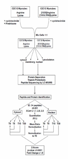
          De novo translation of proteins was inhibited in both SILAC labeled and unlabeled C 2C 12 myotubes. Unlabeled cells were exposed to prednisone while SILAC labeled cells remained untreated. After 0, 5, 15, and 30 minutes of prednisone exposure, labeled and unlabeled cells were mixed at 1:1 ratios and fractionated into cytosolic and nuclear fractions. A total of 534 proteins in the cytosol and 626 proteins in the nucleus were identified and quantitated, using 3 or more peptides per protein with peptide based probability ≤ 0.001. We identified significant increases (1.7- to 3.1- fold) in cytoplasmic abundance of 11 ribosomal proteins within 5 minutes of exposure, all of which returned to baseline by 30 min. We hypothesized that these drug-induced acute changes in the subcellular localization of the cell's protein translational machinery could lead to altered translation of quiescent RNAs. To test this, de novo protein synthesis was assayed after 15 minutes of drug exposure. Quantitative fluorography identified 16 2D gel spots showing rapid changes in translation; five of these were identified by MS/MS (pyruvate kinase, annexin A6 isoform A and isoform B, nasopharyngeal epithelium specific protein 1, and isoform 2 of Replication factor C subunit 1), and all showed the 5' terminal oligopyrimidine motifs associated with mRNA sequestration to and from inactive mRNA pools.

          Conclusion

          We describe novel approaches of subcellular proteomic profiling and assessment of acute changes on a minute-based time scale. These data expand the current knowledge of acute, non-transcriptional activities of glucocorticoids, including changes in protein subcellular localization, altered translation of quiescent RNA pools, and PKC-mediated cytoskeleton remodeling.

          Related collections

          Most cited references46

          • Record: found
          • Abstract: found
          • Article: not found

          Fluorescent indicators for Ca2+ based on green fluorescent proteins and calmodulin.

          Important Ca2+ signals in the cytosol and organelles are often extremely localized and hard to measure. To overcome this problem we have constructed new fluorescent indicators for Ca2+ that are genetically encoded without cofactors and are targetable to specific intracellular locations. We have dubbed these fluorescent indicators 'cameleons'. They consist of tandem fusions of a blue- or cyan-emitting mutant of the green fluorescent protein (GFP), calmodulin, the calmodulin-binding peptide M13, and an enhanced green- or yellow-emitting GFP. Binding of Ca2+ makes calmodulin wrap around the M13 domain, increasing the fluorescence resonance energy transfer (FRET) between the flanking GFPs. Calmodulin mutations can tune the Ca2+ affinities to measure free Ca2+ concentrations in the range 10(-8) to 10(-2) M. We have visualized free Ca2+ dynamics in the cytosol, nucleus and endoplasmic reticulum of single HeLa cells transfected with complementary DNAs encoding chimaeras bearing appropriate localization signals. Ca2+ concentration in the endoplasmic reticulum of individual cells ranged from 60 to 400 microM at rest, and 1 to 50 microM after Ca2+ mobilization. FRET is also an indicator of the reversible intermolecular association of cyan-GFP-labelled calmodulin with yellow-GFP-labelled M13. Thus FRET between GFP mutants can monitor localized Ca2+ signals and protein heterodimerization in individual live cells.
            Bookmark
            • Record: found
            • Abstract: found
            • Article: not found

            Quantitative phosphoproteomics applied to the yeast pheromone signaling pathway.

            Cellular processes such as proliferation, differentiation, and adaptation to environmental changes are regulated by protein phosphorylation. Development of sensitive and comprehensive analytical methods for determination of protein phosphorylation is therefore a necessity in the pursuit of a detailed molecular view of complex biological processes. We present a quantitative modification-specific proteomic approach that combines stable isotope labeling by amino acids in cell culture (SILAC) for quantitation with IMAC for phosphopeptide enrichment and three stages of mass spectrometry (MS/MS/MS) for identification. This integrated phosphoproteomic technology identified and quantified phosphorylation in key regulator and effector proteins of a prototypical G-protein-coupled receptor signaling pathway, the yeast pheromone response. SILAC encoding of yeast proteomes was achieved by incorporation of [(13)C(6)]arginine and [(13)C(6)]lysine in a double auxotroph yeast strain. Pheromone-treated yeast cells were mixed with SILAC-encoded cells as the control and lysed, and extracted proteins were digested with trypsin. Phosphopeptides were enriched by a combination of strong cation exchange chromatography and IMAC. Phosphopeptide fractions were analyzed by LC-MS using a linear ion trap-Fourier transform ion cyclotron resonance mass spectrometer. MS/MS and neutral loss-directed MS/MS/MS analysis allowed detection and sequencing of phosphopeptides with exceptional accuracy and specificity. Of more than 700 identified phosphopeptides, 139 were differentially regulated at least 2-fold in response to mating pheromone. Among these regulated proteins were components belonging to the mitogen-activated protein kinase signaling pathway and to downstream processes including transcriptional regulation, the establishment of polarized growth, and the regulation of the cell cycle.
              Bookmark
              • Record: found
              • Abstract: found
              • Article: not found

              CART classification of human 5' UTR sequences.

              A nonredundant database of 2312 full-length human 5'-untranslated regions (UTRs) was carefully prepared using state-of-the-art experimental and computational technologies. A comprehensive computational analysis of this data was conducted for characterizing the 5' UTR features. Classification and regression tree (CART) analysis was used to classify the data into three distinct classes. Class I consists of mRNAs that are believed to be poorly translated with long 5' UTRs filled with potential inhibitory features. Class II consists of terminal oligopyrimidine tract (TOP) mRNAs that are regulated in a growth-dependent manner, and class III consists of mRNAs with favorable 5' UTR features that may help efficient translation. The most accurate tree we found has 92.5% classification accuracy as estimated by cross validation. The classification model included the presence of TOP, a secondary structure, 5' UTR length, and the presence of upstream AUGs (uAUGs) as the most relevant variables. The present classification and characterization of the 5' UTRs provide precious information for better understanding the translational regulation of human mRNAs. Furthermore, this database and classification can help people build better computational models for predicting the 5'-terminal exon and separating the 5' UTR from the coding region.
                Bookmark

                Author and article information

                Journal
                Proteome Sci
                Proteome Science
                BioMed Central
                1477-5956
                2009
                30 July 2009
                : 7
                : 26
                Affiliations
                [1 ]Research Center for Genetic Medicine, Children's National Medical Center, 111 Michigan Ave NW, Washington DC. 20010, USA
                Article
                1477-5956-7-26
                10.1186/1477-5956-7-26
                2725035
                19642986
                e816e7da-d9ba-4463-9b0a-a1141d9a713c
                Copyright © 2009 Reeves et al; licensee BioMed Central Ltd.

                This is an Open Access article distributed under the terms of the Creative Commons Attribution License ( http://creativecommons.org/licenses/by/2.0), which permits unrestricted use, distribution, and reproduction in any medium, provided the original work is properly cited.

                History
                : 10 March 2009
                : 30 July 2009
                Categories
                Research

                Molecular biology
                Molecular biology

                Comments

                Comment on this article